Difference between revisions of "Chondroma"
Jump to navigation
Jump to search
m (touch) |
|||
| Line 38: | Line 38: | ||
*Multiple chondromas = ''enchondromatosis''; three distinct syndromes:<ref name=emed_enchondroma>URL: [http://emedicine.medscape.com/article/389224-overview http://emedicine.medscape.com/article/389224-overview]. Accessed on: 25 December 2010.</ref> | *Multiple chondromas = ''enchondromatosis''; three distinct syndromes:<ref name=emed_enchondroma>URL: [http://emedicine.medscape.com/article/389224-overview http://emedicine.medscape.com/article/389224-overview]. Accessed on: 25 December 2010.</ref> | ||
**Ollier disease. | **Ollier disease. | ||
**Maffucci syndrome - with [[hemangioma]]s, increased risk of [[chondrosarcoma]].<ref name=omim166000>{{OMIM|166000}}</ref> | **[[Maffucci syndrome]] - with [[hemangioma]]s, increased risk of [[chondrosarcoma]].<ref name=omim166000>{{OMIM|166000}}</ref> | ||
**Metachondromatosis - autosomal dominant. | **Metachondromatosis - autosomal dominant. | ||
*''Enchondroma'' = chondroma in the marrow space. | *''Enchondroma'' = chondroma in the marrow space. | ||
Latest revision as of 21:16, 16 May 2022
| Chondroma | |
|---|---|
| Diagnosis in short | |
 Enchondroma. H&E stain. | |
|
| |
| LM | cytologically benign cells is spaced nests, should not extend into surrounding soft tissue |
| LM DDx | low-grade chondrosarcoma |
| Site | cartilage - see chondro-osseous tumours |
|
| |
| Syndromes | Ollier disease, Maffucci syndrome, metachondromatosis |
|
| |
| Prognosis | benign |
Chondroma is a benign tumour of cartilage. It is in the chondro-osseous tumours group of soft tissue tumours.
General
- Benign thingy.
- Usual legs and feet.
- May be difficult to separate from chondrosarcoma.
- Multiple chondromas = enchondromatosis; three distinct syndromes:[1]
- Ollier disease.
- Maffucci syndrome - with hemangiomas, increased risk of chondrosarcoma.[2]
- Metachondromatosis - autosomal dominant.
- Enchondroma = chondroma in the marrow space.
Clinical:[1]
- Pain.
Radiology
Features:[1]
- Lytic lesion.
- Usual close to a growth plate.
Important suspicious findings that favour malignant:[3]
- Cortical destruction.
- Soft tissue component.
Note:
- High-grade chondroid lesions (high-grade chondrosarcoma) can usually be separated radiologically from low-grade ones.[4]
Microscopic
Features:
- Cytologically benign cells is spaced nests.
- Should not extending into surrounding soft tissue.
DDx:
- Low-grade chondrosarcoma - should be considered, correlation with radiology essential.
Images
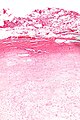
Enchondroma - intermed mag. (WC)
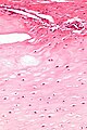
Enchondroma - very high mag. (WC)

Sign out
TISSUE ("CHONDROMA"), LEFT COSTAL MARGIN, EXCISION:
- CHONDROMA.
Micro
The sections show spaced small cells in a pale matrix with a light-blue tinge. No nuclear atypia is appreciated. No mitotic activity is apparent. Degenerative changes are seen focally.
A small focus of cholesterol clefts with giant cells is present. Benign bone, bone marrow and skeletal muscle are present.
See also
References
- ↑ 1.0 1.1 1.2 URL: http://emedicine.medscape.com/article/389224-overview. Accessed on: 25 December 2010.
- ↑ Online 'Mendelian Inheritance in Man' (OMIM) 166000
- ↑ Choi, BB.; Jee, WH.; Sunwoo, HJ.; Cho, JH.; Kim, JY.; Chun, KA.; Hong, SJ.; Chung, HW. et al. "MR differentiation of low-grade chondrosarcoma from enchondroma.". Clin Imaging 37 (3): 542-7. doi:10.1016/j.clinimag.2012.08.006. PMID 23041161.
- ↑ Berber, O.; Datta, G.; Sabharwal, S.; Aston, W.; Saifuddin, A.; Briggs, T. (Apr 2012). "The safety of direct primary excision of low-grade chondral lesions based on radiological diagnosis alone.". Acta Orthop Belg 78 (2): 254-62. PMID 22696998.