Difference between revisions of "Chondroblastoma"
Jump to navigation
Jump to search
(fix) |
(→Images) |
||
| (17 intermediate revisions by 2 users not shown) | |||
| Line 1: | Line 1: | ||
{{ Infobox diagnosis | |||
| Name = {{PAGENAME}} | |||
| Image = Chondroblastoma_-_very_high_mag.jpg | |||
| Width = | |||
| Caption = Chondroblastoma. [[H&E stain]]. | |||
| Synonyms = | |||
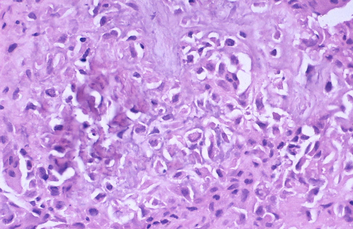
| Micro = abundant (chondroid) extracellular material, chondroblasts (variable nuclear morphology (ovoid, folded or grooved), moderate-abundant eosinophilic cytoplasm), +/-calcifications surrounding the cell nests ("chickenwire" appearance) - classic feature, +/-[[giant cells]] | |||
| Subtypes = | |||
| LMDDx = [[giant cell tumour of bone]], [[chondroma]], well-differentiated [[chondrosarcoma]] | |||
| Stains = | |||
| IHC = | |||
| EM = | |||
| Molecular = | |||
| IF = | |||
| Gross = | |||
| Grossing = | |||
| Site = growth plate - see ''[[chondro-osseous tumours]]'' | |||
| Assdx = | |||
| Syndromes = | |||
| Clinicalhx = "young" - growth plates open | |||
| Signs = | |||
| Symptoms = painful | |||
| Prevalence = | |||
| Bloodwork = | |||
| Rads = | |||
| Endoscopy = | |||
| Prognosis = benign | |||
| Other = | |||
| ClinDDx = | |||
| Tx = | |||
}} | |||
'''Chondroblastoma''' is a benign [[chondro-osseous tumour]] that afflicts the young (growth plates open). | |||
==General== | |||
*Growth plate lesion. | |||
*Sclerotic margin. | |||
*"Young" = growth plates open. | |||
*Most common in teens. | |||
*Typically painful.<ref name=Ref_PCPBoD8_625>{{Ref PCPBoD8|625}}</ref> | |||
*Radiographic osteolytic lesion of the epiphysis [http://radiopaedia.org/articles/lytic-bone-lesion-mnemonic] | |||
*Rare, 1 % of all bone tumors | |||
*Benign | |||
*Humerus, tibia, femur | |||
==Gross== | |||
*Well-defined lesion. | |||
===Image=== | |||
*[http://www.flickr.com/photos/35441329@N05/4052875138/in/set-72157622681280610 Chondroblastoma (flickr.com/humpath)]. | |||
==Microscopic== | |||
Features:<ref name=Ref_WMSP_642>{{Ref WMSP|642}}</ref> | |||
*Abundant extracellular material - pink on [[H&E stain]] - looks vaguely like cartilage. | |||
*Sometimes described as 'immature cartilage' (very narrow DDX for this type of cartilage) | |||
*Chondroblasts: | |||
**Nuclear morphology variable: ovoid, folded or grooved. | |||
**Moderate-abundant eosinophilic cytoplasm. | |||
*+/-Calcification surrounds the cell nests ("chickenwire" appearance) - '''classic feature'''. | |||
**Cell nests have a thin pale blue rimming. | |||
*+/-[[Giant cells]]. | |||
**May lead to confusion with ''[[giant cell tumour of bone]]''. | |||
**Not infrequently associated with an aneurysmal bone cyst (33%).<ref>{{Cite journal | last1 = Sepah | first1 = YJ. | last2 = Umer | first2 = M. | last3 = Minhas | first3 = K. | last4 = Hafeez | first4 = K. | title = Chondroblastoma of the cuboid with an associated aneurysmal bone cyst: a case report. | journal = J Med Case Rep | volume = 1 | issue = | pages = 135 | month = | year = 2007 | doi = 10.1186/1752-1947-1-135 | PMID = 17999776 }}</ref> | |||
DDx: | |||
*[[Giant cell tumour of bone]]. | |||
*[[Chondroma]]. | |||
*Well-differentiated [[chondrosarcoma]]. | |||
*Chondromyxoid fibroma - also has 'immature cartilage' | |||
*[[Aneurysmal bone cyst]] - dont forget that these may be secondary to another lesion. | |||
===Images=== | |||
<gallery> | |||
Image:Chondroblastoma_-_intermed_mag.jpg | Chondroblastoma - intermed. mag. (WC) | |||
Image:Chondroblastoma_-_very_high_mag.jpg | Chondroblastoma - very high mag. (WC) | |||
Image:Bone Chondroblastoma Medium Power.jpg|Chicken wire calcification. (SKB) | |||
Image:Bone Chondroblastoma High Power.jpg|Chicken wire calcification. (SKB) | |||
Image:Bone Chondroblastoma and Aneurysmal Bone Cyst Low Power.jpg|Immature cartilage surrounded by aneurysmal bone cyst with giant cells. (SKB) | |||
Image:Bone Chondroblastoma With Aneurysmal Bone Cyst - Medium Power.jpg|Immature cartilage (left) abutting aneurysmal bone cyst (right) with giant cells. (SKB) | |||
Image:Bone Chondroblastoma - Medium Power.jpg|Immature cartilage. (SKB) | |||
</gallery> | |||
www: | |||
*[http://img.medscape.com/pi/emed/ckb/orthopedic_surgery/1230552-1254949-996.jpg Chondroblastoma (medscape.com)].<ref name=emed_chondroblastoma>URL: [http://emedicine.medscape.com/article/1254949-diagnosis http://emedicine.medscape.com/article/1254949-diagnosis]. Accessed on: 31 December 2010.</ref> | |||
*[http://img.medscape.com/pi/emed/ckb/orthopedic_surgery/1230552-1254949-997.jpg Chondroblastoma with "chickenwire" appearance (medscape.com)].<ref name=emed_chondroblastoma>URL: [http://emedicine.medscape.com/article/1254949-diagnosis http://emedicine.medscape.com/article/1254949-diagnosis]. Accessed on: 31 December 2010.</ref> | |||
*[http://path.upmc.edu/cases/case494/images/fig2a.jpg Chondroblastoma (upmc.edu)].<ref>URL: [http://path.upmc.edu/cases/case494.html http://path.upmc.edu/cases/case494.html]. Accessed on: 24 January 2012.</ref> | |||
*Tumor Library - case with giant cells[http://www.tumorlibrary.com/case/image.jsp?title=Chondroblastoma+-+Distal+femur+-+Photomicrograph&uri=/case/images/3571.jpg] | |||
==IHC== | |||
Features:<ref name=Ref_WMSP_642>{{Ref WMSP|642}}</ref> | |||
*S100 +ve. | |||
*Vimentin +ve.<ref name=emed_chondroblastoma>URL: [http://emedicine.medscape.com/article/1254949-diagnosis http://emedicine.medscape.com/article/1254949-diagnosis]. Accessed on: 31 December 2010.</ref> | |||
==See also== | |||
*[[Chondro-osseous tumours]]. | |||
*Tumor Library[http://www.tumorlibrary.com/case/list.jsp?order=diagnosis+ASC&diagnosis=Chondroblastoma] | |||
==References== | |||
{{Reflist|2}} | |||
[[Category:Diagnosis]] | |||
[[Category:Chondro-osseous tumours]] | |||
Latest revision as of 05:24, 21 October 2014
| Chondroblastoma | |
|---|---|
| Diagnosis in short | |
 Chondroblastoma. H&E stain. | |
|
| |
| LM | abundant (chondroid) extracellular material, chondroblasts (variable nuclear morphology (ovoid, folded or grooved), moderate-abundant eosinophilic cytoplasm), +/-calcifications surrounding the cell nests ("chickenwire" appearance) - classic feature, +/-giant cells |
| LM DDx | giant cell tumour of bone, chondroma, well-differentiated chondrosarcoma |
| Site | growth plate - see chondro-osseous tumours |
|
| |
| Clinical history | "young" - growth plates open |
| Symptoms | painful |
| Prognosis | benign |
Chondroblastoma is a benign chondro-osseous tumour that afflicts the young (growth plates open).
General
- Growth plate lesion.
- Sclerotic margin.
- "Young" = growth plates open.
- Most common in teens.
- Typically painful.[1]
- Radiographic osteolytic lesion of the epiphysis [1]
- Rare, 1 % of all bone tumors
- Benign
- Humerus, tibia, femur
Gross
- Well-defined lesion.
Image
Microscopic
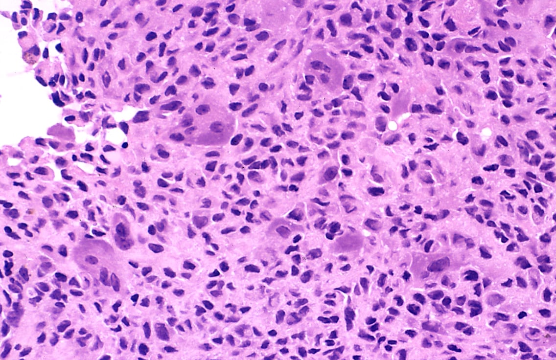
Features:[2]
- Abundant extracellular material - pink on H&E stain - looks vaguely like cartilage.
- Sometimes described as 'immature cartilage' (very narrow DDX for this type of cartilage)
- Chondroblasts:
- Nuclear morphology variable: ovoid, folded or grooved.
- Moderate-abundant eosinophilic cytoplasm.
- +/-Calcification surrounds the cell nests ("chickenwire" appearance) - classic feature.
- Cell nests have a thin pale blue rimming.
- +/-Giant cells.
- May lead to confusion with giant cell tumour of bone.
- Not infrequently associated with an aneurysmal bone cyst (33%).[3]
DDx:
- Giant cell tumour of bone.
- Chondroma.
- Well-differentiated chondrosarcoma.
- Chondromyxoid fibroma - also has 'immature cartilage'
- Aneurysmal bone cyst - dont forget that these may be secondary to another lesion.
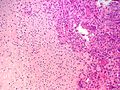
Images
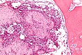
Chondroblastoma - intermed. mag. (WC)
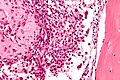
Chondroblastoma - very high mag. (WC)
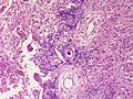
Chicken wire calcification. (SKB)
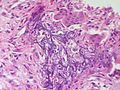
Chicken wire calcification. (SKB)
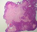
Immature cartilage surrounded by aneurysmal bone cyst with giant cells. (SKB)
Immature cartilage (left) abutting aneurysmal bone cyst (right) with giant cells. (SKB)
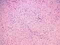
Immature cartilage. (SKB)


www:
- Chondroblastoma (medscape.com).[4]
- Chondroblastoma with "chickenwire" appearance (medscape.com).[4]
- Chondroblastoma (upmc.edu).[5]
- Tumor Library - case with giant cells[2]
![[2]](http://www.tumorlibrary.com/case/image.jsp?title=Chondroblastoma+-+Distal+femur+-+Photomicrograph&uri=/case/images/3571.jpg)
IHC
Features:[2]
- S100 +ve.
- Vimentin +ve.[4]
See also
- Chondro-osseous tumours.
- Tumor Library[3]
References
- ↑ Mitchell, Richard; Kumar, Vinay; Fausto, Nelson; Abbas, Abul K.; Aster, Jon (2011). Pocket Companion to Robbins & Cotran Pathologic Basis of Disease (8th ed.). Elsevier Saunders. pp. 625. ISBN 978-1416054542.
- ↑ 2.0 2.1 Humphrey, Peter A; Dehner, Louis P; Pfeifer, John D (2008). The Washington Manual of Surgical Pathology (1st ed.). Lippincott Williams & Wilkins. pp. 642. ISBN 978-0781765275.
- ↑ Sepah, YJ.; Umer, M.; Minhas, K.; Hafeez, K. (2007). "Chondroblastoma of the cuboid with an associated aneurysmal bone cyst: a case report.". J Med Case Rep 1: 135. doi:10.1186/1752-1947-1-135. PMID 17999776.
- ↑ 4.0 4.1 4.2 URL: http://emedicine.medscape.com/article/1254949-diagnosis. Accessed on: 31 December 2010.
- ↑ URL: http://path.upmc.edu/cases/case494.html. Accessed on: 24 January 2012.