Breast grossing
Jump to navigation
Jump to search
The printable version is no longer supported and may have rendering errors. Please update your browser bookmarks and please use the default browser print function instead.

.jpg)
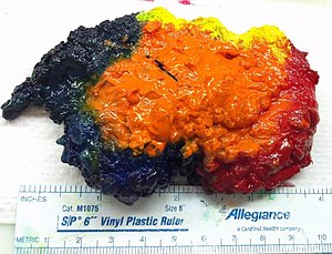
An inked breast lumpectomy specimen. (WC/Ed Uthman)
This article deals with breast grossing.
Introduction
- Lumpectomy = a common procedure for breast lesions that are small, typically have no skin.
- Mastectomy = removal of the breast, may include skeletal muscle (not common) or be skin sparing.[1]
Specimen opening
- Orientation:
- Lumpectomies are usually oriented with a short and long suture as per the surgeon; short is typically superior (aspect) and long is typically lateral (aspect).
- Mastectomies typically have tissue extending toward the axilla known as the "tail".
- The deep aspect in larger specimens can often be identified by the (flat) fascial plane.
- Inking - colours usually as per an institutional standard - see Protocol notes section.
- Slicing - medial to lateral.
Protocol
Identification:
- Specimen label: [description as per label].
- Specimen label and requisition: [match/do not match].
Specimen - type/size/characteristics:
- Specimen type: [total mastectomy/partial mastectomy].
- Specimen orientation: [short-superior, long-lateral, double deep].
- Surgical guidewire: [present/absent].
- Specimen size (superior-inferior, medial-lateral, anterior-posterior): [___ x ___ x ___] cm.
- Surface disruption/intactness: [intact/disrupted at (location) - defect measures ___ cm].
- Skin: [___ x ___ cm/absent].
- Axilla: [___ x ___ x ___ cm, [mass lesion ___x___x___ cm/mass lesion absent]/axillary tissue absent].
- Nipple: [___ length cm x ___ diameter cm, [unremarkable appearance/retracted]/nipple absent].
- Skeletal muscle: [present, [unremarkable appearance/fibrotic/suspicious for tumour/involved by tumour]/skeletal muscle absent].
- Inking code: [posterior-black, anterior-yellow, superior-blue, interior-red].‡
Slices:
- Slicing: [medial-to-lateral, parasagittal cuts].
- Number of slices: [number].
- Slices sent to x-ray: [yes/no].
- Calcifications: [present/not identified].
Tumour:
- Tumour location in slices: [___ to ___].
- Tumour size (superior-inferior, medial-lateral, anterior-posterior): [___ x ___ x ___] cm.
- Closest margin and distance: [___ margin, ___ cm].
- Distance to other margins: [anterior: [___ cm/not applicable], posterior: [___ cm/not applicable], superior [___ cm/not applicable], inferior [___ cm/not applicable], medial: [___ cm/not applicable], lateral: [___ cm/not applicable].
Other:
- Uninvolved parenchyma - appearance: [fibrous/fatty].
- Other findings: [none/description of other findings].
Sections:
- Margins - on edge if section can be taken with tumour and margin.
- Tumour - in total if small (<2 cm[2]).
Protocol notes
- ‡ There is no universally accepted inking protocol. Blue for superior and green for inferior is common, as the sky is blue and the grass is green.
- Hua[2] suggests: black = posterior, blue = superior, green = inferior, yellow = anterior, red = medial & lateral.
Staging
Main article: Breast cancer staging
The important cut-points (at the time of gross) for tumour staging are: 5, 10, 20, 50 mm.
Alternate approaches
See also
Related protocols
References
- ↑ Yu, P. (Aug 2016). "Breast reconstruction at the MD Anderson Cancer Center.". Gland Surg 5 (4): 416-21. doi:10.21037/gs.2016.05.03. PMID 27563563.
- ↑ 2.0 2.1 Huo, L. (Aug 2011). "A practical approach to grossing breast specimens.". Ann Diagn Pathol 15 (4): 291-301. doi:10.1016/j.anndiagpath.2011.03.005. PMID 21745648.