Benign cortical cyst of the kidney
Jump to navigation
Jump to search
| Benign cortical cyst of the kidney | |
|---|---|
| Diagnosis in short | |
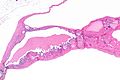
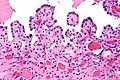
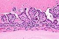
 Benign cortical renal cyst with papillary projections. H&E stain. | |
|
| |
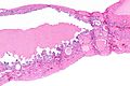
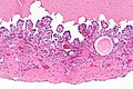
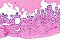
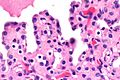
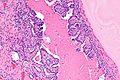
| LM | simple epithelial lining without atypia, may have clear cells |
| LM DDx | localized cystic disease of the kidney, cystic neoplasms/renal cell carcinoma (e.g. cystic clear cell renal cell carcinoma), multicystic renal cell neoplasm of low malignant potential, other cystic kidney diseases |
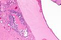
| Gross | thin-walled cyst in the renal cortex, usually filled with serous fluid, usually unilocular |
| Site | kidney - see cystic kidney diseases |
|
| |
| Prevalence | very common |
| Radiology | simple cyst with thin wall; usually Bosniak I or II - see Bosniak classification of renal cysts |
| Prognosis | benign |
| Treatment | none |
Benign cortical cyst of the kidney is an extremely common benign kidney finding.
Benign renal cyst redirects here. A more general discussion about cysts in the kidney is in the article cystic kidney diseases.
General
- Very common.
- Benign.
Gross
- Thin-walled cyst - usually filled with serous fluid.
- Usually unilocular.
Microscopic
Features:
- Simple epithelial lining without atypia.
- May have clear cells.
Note:
- Do not have clear cells within the wall of the cyst.[1]
DDx:
- Localized cystic disease of the kidney.
- Cystic neoplasms/renal cell carcinoma:
- Cystic clear cell renal cell carcinoma - thick septa with clear cells.
- Multicystic renal cell neoplasm of low malignant potential - thin septa, clear cells within stroma.
- Other cystic kidney diseases.
Images
ARC - very low mag. (WC)
ARC - low mag. (WC)
ARC - intermed. mag. (WC)
ARC - intermed. mag. (WC)
ARC - high mag. (WC)
ARC - very high mag. (WC)
ARC - low mag. (WC)
ARC - intermed. mag. (WC)
ARC - high mag. (WC)


See also
References
- ↑ Epstein, Jonathan I.; Netto, George J. (2014). Differential Diagnoses in Surgical Pathology: Genitourinary System (1st ed.). Wolters Kluwer. pp. 197. ISBN 978-1451189582.