Bacteria
Bacteria are single celled microorganisms, without a nucleus (prokaryotes), that can cause lots of morbidity and mortality. They are not infrequently seen by pathologists.
Actinobacteria
General
- A very large group of bacteria.
It includes:[1]
- Actinomycetes.
- Corynebacterium.
- Mycobacterium.
- Nocardia.
- Streptomyces.
Actinomycetes
General
- IUD needs to be removed if found on a pap test[2] - see gynecologic cytopathology.
- Gram-positive branching rods.
- Common in the tonsils.
- Part of the large Actinobacteria group.[1]
Notes:
- Mycete = fungus; these organisms have a fungus-like appearance.
- Also called pseudomycosis.
Gross
- Yellow granules.[3]
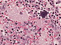
Microscopic
- Branching rods.
- Typically form pink/purple granules ("sulfur granule") that is surrounded by inflammatory cells (lymphocytes or neutrophils).
DDx:
- Nocardia.
Notes:
- Sulfur granule may be seen grossly - yellow.[3]
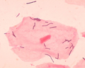
Images
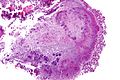
Actinomyces - high mag. (WC)
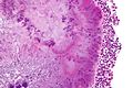
Actinomyces - very high mag. (WC)
Purulent encephalitis with actinomyces in HE. (WC/jensflorian)
Purulent encephalitis with actinomyces in Grocott. (WC/jensflorian)


Stains
- Gram +ve.
- AFB -ve.
- Nocardia +ve.
- PAS +ve.
- May be confused with a fungus!
- Grocott +ve.
Helicobacter pylori
- Commonly abbreviated H. pylori or HP.
General
- Gram-negative rods.[5]
- Causes gastritis - specifically Helicobacter gastritis.
- Associated with peptic ulcer disease, MALT lymphoma and gastric carcinoma.
Microscopy
Clostridium difficile
- Commonly C. difficile.
- Classic cause of pseudomembranous colitis.
General
- Gram positive bacillus.
One virulent strain is:[6][7] BI/NAP1.
- Deletion of tcdC locus.
- Resistant to fluoroquinolones (gatifloxacin and moxifloxacin).
Chlamydia trachomatis
- May be referred to as Chlamydia.
General
- Common.
- May cause lymphogranuloma venereum.
Note:
- Often co-exists with gonorrhea.
Microscopic
- Variable.
Lymphogranuloma venereum:
- See: Cat-scratch disease.
Mycobacterium tuberculosis
- Abbreviated TB.
General
- Causes tuberculosis.
- May mimic a malignancy.
- Strong association with HIV.
- TB has characteristics of Gram positive and Gram negative bacteria.[8]
Clinical
Classic features - pulmonary/systemic:
- Cough.
- Fever.
- Weight loss.
CNS manifestations:
- Tuberculoma (mass).
- Meningitis.
- Abscess.
Tests:
Treatment:
- Multiple drugs for a long time (months).
- Commonly used drugs: isoniazid, rifampin, pyrazinamide, and ethambutol.
Gross
Ghon complex
Consists of two components:[12][13]
- Peripheral focus - subpleural, calcified.
- Central focus - the hilar lymph node that drains the peripheral focus.
Image:
Microscopic
Features:
- Necrotizing granulomas with rod-shaped bacteria.
Note:
- May be non-necrotizing.
DDx:
Images:
- Tuberculosis - case 1 - several images (upmc.edu).
- Tuberculosis - case 2 - several images (upmc.edu).
- Tuberculosis - case 3 - several images (upmc.edu).
Stains
- Ziehl-Neelsen stain - red rod-shaped bacteria - key feature.
- Very small - must use 40x objective.
Image:
Molecular
- Can be diagnosed with PCR.
Mycobacterium leprae
General
- Causes leprosy.
Clinical:
- Nerve damage -> injuries -> disability.
Microscopic
Features:
- Granulomas with rod-shaped bacteria.
Stains
- Fite stain - red rod-shaped bacteria - key feature.
- Very small - must use 40x objective.
Images:
Mycobacterium avium complex
- Abbreviated MAC.
- Previously referred to as Mycobacterium avium-intracellulare, abbreviated MAI.
General
- Refers to an infection with both:[16]
- Mycobacterium avium
- Mycobacterium intracellulare.
Microscopic
Features:
- Small rod-shaped organisms - within histocytes.
- +/-Granulomas.
DDx:
- Tuberculosis.
- Whipple disease - esp. in the duodenum.
Stains
- AFB +ve.
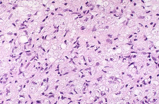
Coxiella burnetii
General
- Causes: Q fever.
Features:
- Intracellular bacterium.
- Gram negative.
Clinical:
- Flu-like symptoms.
Microscopic
Features:
- Fibrin ring granuloma.
- Epithelioid macrophages (i.e. a granuloma) surrounding a thin pink (fibrin) ring.
DDx:[17]
- Infections (Coxiella burnetii, CMV, EBV + others).
- Drug reaction.
- Malignancy (e.g. Hodgkin lymphoma[18]).
Images:
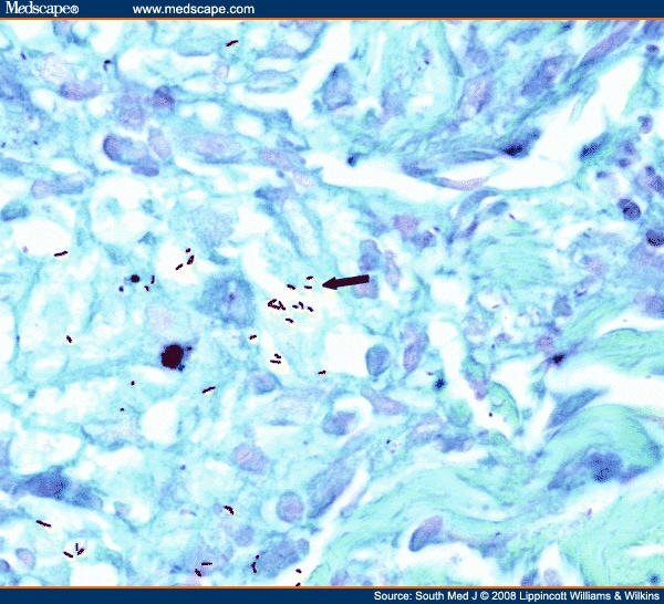
Bartonella henselae
General
Causative agent in:
Microscopic
Features - bacillary angiomatosis:
- Similar to pyogenic granuloma - see pyogenic granuloma.
Features - cat-scratch disease:
- Stellate granulomas.
Stains
- Warthin-Starry stain +ve.
Image:
Lactobacillus
General
- Gram positive bacilli.
- Normal vaginal flora.
Microscopic
Features:
- Slender bacilli.
Lactobacilli. (WC)

Pseudomonas
General
- Gram-negative bacteria.
- Common pathogenic Pseudomonas aeruginosa.
- Community-acquired bronchopneumonia.
Gross
- Green nail.
Images:
Sarcina
See also
References
- ↑ 1.0 1.1 Ventura, M.; Canchaya, C.; Tauch, A.; Chandra, G.; Fitzgerald, GF.; Chater, KF.; van Sinderen, D. (Sep 2007). "Genomics of Actinobacteria: tracing the evolutionary history of an ancient phylum.". Microbiol Mol Biol Rev 71 (3): 495-548. doi:10.1128/MMBR.00005-07. PMC 2168647. PMID 17804669. https://www.ncbi.nlm.nih.gov/pmc/articles/PMC2168647/.
- ↑ Humphrey, Peter A; Dehner, Louis P; Pfeifer, John D (2008). The Washington Manual of Surgical Pathology (1st ed.). Lippincott Williams & Wilkins. pp. 446. ISBN 978-0781765275.
- ↑ 3.0 3.1 3.2 URL: http://pathmicro.med.sc.edu/mycology/mycology-2.htm. Accessed on: 14 September 2011.
- ↑ URL: http://pathology.class.kmu.edu.tw/ch05/Slide42.htm . Accessed on: 14 September 2011.
- ↑ Mobley, HLT.; Mendz, GL.; Hazell, SL.; Andersen, LP.; Wadström, T.. Basic Bacteriology and Culture. PMID 21290743. http://www.ncbi.nlm.nih.gov/books/NBK2444/.
- ↑ URL: http://www.medpagetoday.com/InfectiousDisease/PublicHealth/2254. Accessed on: 15 August 2011.
- ↑ McDonald, LC.; Killgore, GE.; Thompson, A.; Owens, RC.; Kazakova, SV.; Sambol, SP.; Johnson, S.; Gerding, DN. (Dec 2005). "An epidemic, toxin gene-variant strain of Clostridium difficile.". N Engl J Med 353 (23): 2433-41. doi:10.1056/NEJMoa051590. PMID 16322603.
- ↑ Fu, LM.; Fu-Liu, CS. (2002). "Is Mycobacterium tuberculosis a closer relative to Gram-positive or Gram-negative bacterial pathogens?". Tuberculosis (Edinb) 82 (2-3): 85-90. PMID 12356459.
- ↑ Kawakami, S.; Kawamura, Y.; Nishiyama, K.; Hatanaka, H.; Fujisaki, R.; Ono, Y.; Miyazawa, Y.; Nishiya, H. (Dec 2012). "Case of Mycobacterium tuberculosis meningitis: Gram staining as a useful initial diagnostic clue for tuberculous meningitis.". J Infect Chemother 18 (6): 931-6. doi:10.1007/s10156-012-0382-y. PMID 22476652.
- ↑ Atsukawa, Y.; Kawakami, S.; Asahara, M.; Ishigaki, S.; Tanaka, T.; Ono, Y.; Nishiya, H.; Fujisaki, R. et al. (Aug 2011). "The usefulness of changing focus during examination using Gram staining as initial diagnostic clue for infective tuberculosis.". J Infect Chemother 17 (4): 571-4. doi:10.1007/s10156-011-0216-3. PMID 21327691.
- ↑ Fu, LM.; Fu-Liu, CS. (2002). "Genome comparison of Mycobacterium tuberculosis and other bacteria.". OMICS 6 (2): 199-206. doi:10.1089/153623102760092797. PMID 12143965.
- ↑ Rose, Alan G. (2008). Atlas of Gross Pathology with Histologic Correlation (1st ed.). Cambridge University Press. pp. 112. ISBN 978-0521868792.
- ↑ URL: http://pathhsw5m54.ucsf.edu/case32/image324.html. Accessed on: 27 February 2012.
- ↑ URL: http://www.medscape.com/viewarticle/576467_2. Accessed on: 2 January 2012.
- ↑ URL: http://www.meddean.luc.edu/lumen/MedEd/orfpath/bfsrinf.htm. Accessed on: 1 April 2012.
- ↑ Turenne, CY.; Wallace, R.; Behr, MA. (Apr 2007). "Mycobacterium avium in the postgenomic era.". Clin Microbiol Rev 20 (2): 205-29. doi:10.1128/CMR.00036-06. PMC 1865596. PMID 17428883. https://www.ncbi.nlm.nih.gov/pmc/articles/PMC1865596/.
- ↑ Tjwa M, De Hertogh G, Neuville B, Roskams T, Nevens F, Van Steenbergen W (2001). "Hepatic fibrin-ring granulomas in granulomatous hepatitis: report of four cases and review of the literature". Acta Clin Belg 56 (6): 341–8. PMID 11881318.
- ↑ de Bayser L, Roblot P, Ramassamy A, Silvain C, Levillain P, Becq-Giraudon B (July 1993). "Hepatic fibrin-ring granulomas in giant cell arteritis". Gastroenterology 105 (1): 272–3. PMID 8514044.
- ↑ 19.0 19.1 Barankin, B.; Levy, J. (Oct 2012). "Dermacase. Can you identify this condition? Pseudomonas aeruginosa infection.". Can Fam Physician 58 (10): 1103-4. PMID 23064921.
- ↑ Hengge, UR.; Bardeli, V. (Mar 2009). "Images in clinical medicine. Green nails.". N Engl J Med 360 (11): 1125. doi:10.1056/NEJMicm0706497. PMID 19279344.