Difference between revisions of "Autoimmune metaplastic atrophic gastritis"
(→Images: sm fix) |
|||
| (2 intermediate revisions by the same user not shown) | |||
| Line 38: | Line 38: | ||
*Parietal cells. | *Parietal cells. | ||
*Intrinsic factor. | *Intrinsic factor. | ||
Others: | Others: | ||
*Gastrin level (increased).<ref name=pmid21947876>{{Cite journal | last1 = Annibale | first1 = B. | last2 = Lahner | first2 = E. | last3 = Fave | first3 = GD. | title = Diagnosis and management of pernicious anemia. | journal = Curr Gastroenterol Rep | volume = 13 | issue = 6 | pages = 518-24 | month = Dec | year = 2011 | doi = 10.1007/s11894-011-0225-5 | PMID = 21947876 }}</ref> | *Gastrin level (increased).<ref name=pmid21947876>{{Cite journal | last1 = Annibale | first1 = B. | last2 = Lahner | first2 = E. | last3 = Fave | first3 = GD. | title = Diagnosis and management of pernicious anemia. | journal = Curr Gastroenterol Rep | volume = 13 | issue = 6 | pages = 518-24 | month = Dec | year = 2011 | doi = 10.1007/s11894-011-0225-5 | PMID = 21947876 }}</ref> | ||
| Line 62: | Line 62: | ||
*[[Gastric neuroendocrine tumour]]. | *[[Gastric neuroendocrine tumour]]. | ||
*[[Intestinal metaplasia of the stomach]] with chronic inflammation. | *[[Intestinal metaplasia of the stomach]] with chronic inflammation. | ||
*[[Chronic gastritis]]. | |||
===Images=== | ===Images=== | ||
| Line 118: | Line 119: | ||
==Sign out== | ==Sign out== | ||
<pre> | |||
Stomach, Body, Biopsy: | |||
- Gastric mucosa with INTESTINAL METAPLASIA, | |||
moderate chronic inactive inflammation and atrophic features, SEE COMMENT. | |||
- NEGATIVE for apparent parietal cells, SEE COMMENT. | |||
- NEGATIVE for Helicobacter-like organisms. | |||
- NEGATIVE for dysplasia and NEGATIVE for malignancy. | |||
Comment: | |||
Immunostains show rows of Chromogranin A positive cells and a lack of gastrin staining. | |||
These findings suggest an autoimmune (metaplastic atrophic) gastritis; correlation with blood work and clinical findings is recommended. | |||
</pre> | |||
===Block letters=== | |||
<pre> | <pre> | ||
STOMACH, BIOPSY: | STOMACH, BIOPSY: | ||
Latest revision as of 00:08, 26 March 2024
| Autoimmune metaplastic atrophic gastritis | |
|---|---|
| Diagnosis in short | |
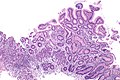
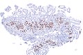
 Atrophic gastritis (body) without appreciable parietal cells. H&E stain. | |
|
| |
| LM | corpus predominant inflammation - usu. moderate or severe, loss of parietal cells, increased G cells in the antrum |
| LM DDx | chronic gastritis, intestinal metaplasia of the stomach, gastric neuroendocrine tumour |
| Site | stomach |
|
| |
| Prevalence | uncommon |
| Blood work | antibodies to parietal cells & intrinsic factor, macrocytic anemia, increased gastrin level |
| Endoscopy | erythema - corpus only |
| Clin. DDx | diffuse chronic gastritis |
Autoimmune metaplastic atrophic gastritis, also autoimmune gastritis[1] (abbreviated AIG), is a rare pathology of the stomach. It is closely associated with pernicious anemia.
General
- Pathology: loss of parietal cells, gastric atrophy.
- Lab: classically considered to have macrocytic anemia; however, normocytic and microcystic more common.[2]
- Etiology: autoimmune.
Diagnosis based on serology for antibodies to:[3]
- Parietal cells.
- Intrinsic factor.
Others:
Note:
- Parietal cells produce intrinsic factor (important for vitamin B12 absorption) and hydrogen chloride, i.e. stomach acid.
Gross
- Erythema - corpus involved, antrum spared.
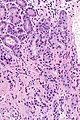
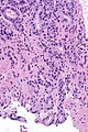
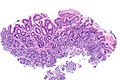
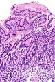
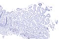
Microscopic
Features:
- Corpus predominant inflammation - usu. moderate or severe - key feature.
- Loss of parietal cells.
- Increased G cells in the antrum.
- Produce gastrin to stimulate the (missing) parietal cells.
Notes:
- Compare with other types of gastric atrophy.
DDx:
- Gastric neuroendocrine tumour.
- Intestinal metaplasia of the stomach with chronic inflammation.
- Chronic gastritis.
Images

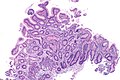
MAG (body) - very low mag.
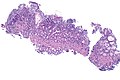
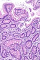
MAG (body) - low mag.
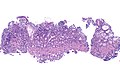
MAG (body) - low mag.
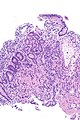
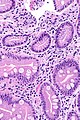
MAG (body) - intermed. mag.
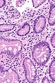
MAG (body) - high mag.
MAG (body) - high mag.
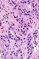
MAG (body) - very high mag.
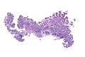
MAG (body) - very low mag.
MAG (body) - low mag.
MAG (body) - low mag.
MAG (body) - intermed. mag.
MAG (body) - high mag.
MAG (body) - high mag.
MAG - antrum - low mag.
MAG - antrum - low mag.
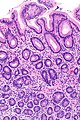
MAG - antrum - low mag.


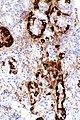
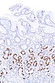
IHC
Features:[6]
- Chromogranin A +ve (demonstrates nodular enterochromaffin-like cell hyperplasia).
- Gastrin -ve (body of stomach).
- +ve in antrum.
Images
Body

MAG (body) - chromogranin A - very low mag.
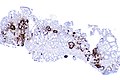
MAG (body) - chromogranin A - low mag.
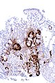
MAG (body) - chromogranin A - intermed. mag.
MAG (body) - chromogranin A - high mag.

MAG (body) - gastrin - low mag.
MAG (body) - gastrin - low mag.


Antrum
MAG (antrum) - gastrin - low mag.
MAG (antrum) - gastrin - intermed. mag.


www
- Autoimmune gastritis - chromogranin A (nih.gov).[7]
- Findings may be seen in hypergastrinemia and nodular enterochromaffin cell-like (ECL) hyperplasia.
Sign out
Stomach, Body, Biopsy:
     - Gastric mucosa with INTESTINAL METAPLASIA,
       moderate chronic inactive inflammation and atrophic features, SEE COMMENT.
     - NEGATIVE for apparent parietal cells, SEE COMMENT.
     - NEGATIVE for Helicobacter-like organisms.
     - NEGATIVE for dysplasia and NEGATIVE for malignancy.

Comment:
Immunostains show rows of Chromogranin A positive cells and a lack of gastrin staining.

These findings suggest an autoimmune (metaplastic atrophic) gastritis; correlation with blood work and clinical findings is recommended.
Block letters
STOMACH, BIOPSY:
- SEVERE CHRONIC ACTIVE GASTRITIS WITH EXTENSIVE INTESTINAL METAPLASIA.
- NEGATIVE FOR HELICOBACTER-LIKE ORGANISMS.
- NEGATIVE FOR DYSPLASIA AND NEGATIVE FOR MALIGNANCY.

COMMENT:
Parietal cells are not apparent on the H&E stained sections. Immunostains show
rows of Chromogranin A positive cells and a lack of gastrin staining.

These findings suggest an autoimmune gastritis; correlation with blood work
is suggested.
See also
References
- ↑ Chlumská, A.; Boudová, L.; Benes, Z.; Zámecník, M. (Oct 2005). "Autoimmune gastritis. A clinicopathologic study of 25 cases.". Cesk Patol 41 (4): 137-42. PMID 16382988.
- ↑ Hershko, C.; Ronson, A.; Souroujon, M.; Maschler, I.; Heyd, J.; Patz, J. (Feb 2006). "Variable hematologic presentation of autoimmune gastritis: age-related progression from iron deficiency to cobalamin depletion.". Blood 107 (4): 1673-9. doi:10.1182/blood-2005-09-3534. PMID 16239424.
- ↑ Oh, R.; Brown, DL. (Mar 2003). "Vitamin B12 deficiency.". Am Fam Physician 67 (5): 979-86. PMID 12643357.
- ↑ Annibale, B.; Lahner, E.; Fave, GD. (Dec 2011). "Diagnosis and management of pernicious anemia.". Curr Gastroenterol Rep 13 (6): 518-24. doi:10.1007/s11894-011-0225-5. PMID 21947876.
- ↑ URL: http://www.mayomedicallaboratories.com/test-catalog/Clinical+and+Interpretive/8512. Accessed on: 14 August 2012.
- ↑ Park, JY.; Cornish, TC.; Lam-Himlin, D.; Shi, C.; Montgomery, E. (Nov 2010). "Gastric lesions in patients with autoimmune metaplastic atrophic gastritis (AMAG) in a tertiary care setting.". Am J Surg Pathol 34 (11): 1591-8. doi:10.1097/PAS.0b013e3181f623af. PMID 20975338.
- ↑ Pritchard, DM.; Berry, D.; Przemeck, SM.; Campbell, F.; Edwards, SW.; Varro, A. (Oct 2008). "Gastrin increases mcl-1 expression in type I gastric carcinoid tumors and a gastric epithelial cell line that expresses the CCK-2 receptor.". Am J Physiol Gastrointest Liver Physiol 295 (4): G798-805. doi:10.1152/ajpgi.00015.2008. PMID 18719002.