Auer rod
Jump to navigation
Jump to search
The printable version is no longer supported and may have rendering errors. Please update your browser bookmarks and please use the default browser print function instead.
.jpg)
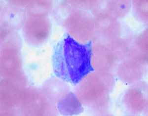
Micrograph showing an Auer rod in a blast. Wright stain. (WC)
Auer rod a microscopic finding typical of some types of acute myeloid leukemia and seen in myelodysplastic syndromes.
It may be referred to as an Auer body.
General
Microscopic
Features:
- Needle-like cytoplasmic bodies - classic appearance.[3]
- Other shapes: comma-like, diamond-like, rectangular, rarely glandular or corkscrew-like.
DDx:
- Auer-rod like inclusions - may be seen in multiple myeloma[4] or lymphoma cells.[2]
Images
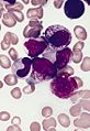
Auer rods. (WC)
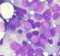
Abundant Auer rods in a Faggot cell. (WC)


See als
References
- ↑ Yoshida, Y.; Oguma, S.; Ohno, H.; Auer, J. (May 2009). "John Auer and Auer rods; controversies revisited.". Leuk Res 33 (5): 614-6. doi:10.1016/j.leukres.2008.09.014. PMID 18947869.
- ↑ 2.0 2.1 Groom, DA.; Wong, D.; Brynes, RK.; Macaulay, LK. (Jul 1991). "Auer rod-like inclusions in circulating lymphoma cells.". Am J Clin Pathol 96 (1): 111-5. PMID 1712539.
- ↑ ACKERMAN, GA. (Sep 1950). "Microscopic and histochemical studies on the Auer bodies in leukemic cells.". Blood 5 (9): 847-63. PMID 15434012.
- ↑ Abdulsalam, AH.; Bain, BJ. (Mar 2014). "Auer-rod like inclusions in multiple myeloma.". Am J Hematol 89 (3): 338. doi:10.1002/ajh.23648. PMID 24338920.