Angiocentric glioma
Jump to navigation
Jump to search
The printable version is no longer supported and may have rendering errors. Please update your browser bookmarks and please use the default browser print function instead.
| Angiocentric glioma | |
|---|---|
| Diagnosis in short | |
 Angiocentric glioma. H&E stain. | |
| LM DDx | astrocytoma, ependymoma. |
| IHC | GFAP +/-ve, EMA +/-ve. |
| Gross | enlargened gyri |
| Site | brain - usu. grey matter |
|
| |
| Clinical history | epilepsy-associated |
| Prevalence | very rare - no age prevalence |
| Prognosis | good (WHO Grade I) |
Angiocentric glioma, is a WHO grade I glioma. It is super rare.
General
- previously called monomorphic angiocentric glioma or angiocentric neuroepithelial tumour.
- Own entity introduced in the WHO 2007 classification.[1]
- Low-grade glioma - WHO Grade I by definition, but a single recurrence with anaplastic features has been described.[2]
- Classically a non-enhancing, superficial cerebrocortical lesion.
- Associated with epilepsy.
- No association with any tumour syndromes.
Gross
- Usually well-circumscribed.
- Enlargement of cortex possible.
Microscopic
Features:
- Monommorphic, bipolar, spindled cells around blood vessels.
- mimicking ependymal pseudorosettes (DD: ependymoma).
- Solid growth with palisaded arrays possible.
- Low cellularity and rich myxoid background- when compared to classical astrocytomas.
- Mitotic activity may lead to eroneous diagnosis of anaplastic astrocytoma.
- Variably GFAP, EMA and S-100 positive
- No IDH1/2 mutations. [3]
- MIB-1 between 1-5%
DDx of angiocentric glioma (brief):
- Pilomyxoid astrocytoma
- Ependymoma.
- Astrocytoma.
- Isomorphic astrocytoma.
Molecular
Note: Gliomas with MYB/MYBL alteration may histologically resemble diffuse astrocytoma in children but these tumors cluster together with angiocentric glioma suggesting a single tumor entity.[6]
Images
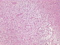
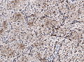
Angiocentric glioma - low mag. (WC/jensflorian)
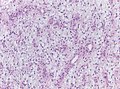
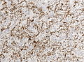
Angiocentric glioma - intermed mag. (WC/jensflorian)
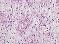
Angiocentric glioma - high mag. (WC/jensflorian)
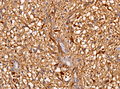
Angiocentric glioma - GFAP immunostain (WC/jensflorian)
Angiocentric glioma - EMA immunostain (WC/jensflorian)
Angiocentric glioma - MAP2 immunostain (WC/jensflorian)
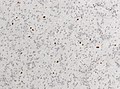
Angiocentric glioma - MIB-1 immunostain (WC/jensflorian)


See also
References
- ↑ Brat, DJ.; Scheithauer, BW.; Fuller, GN.; Tihan, T. (Jul 2007). "Newly codified glial neoplasms of the 2007 WHO Classification of Tumours of the Central Nervous System: angiocentric glioma, pilomyxoid astrocytoma and pituicytoma.". Brain Pathol 17 (3): 319-24. doi:10.1111/j.1750-3639.2007.00082.x. PMID 17598825.
- ↑ Wang, M.; Tihan, T.; Rojiani, AM.; Bodhireddy, SR.; Prayson, RA.; Iacuone, JJ.; Alles, AJ.; Donahue, DJ. et al. (Oct 2005). "Monomorphous angiocentric glioma: a distinctive epileptogenic neoplasm with features of infiltrating astrocytoma and ependymoma.". J Neuropathol Exp Neurol 64 (10): 875-81. PMID 16215459.
- ↑ Raghunathan, A.; Olar, A.; Vogel, H.; Parker, JR.; Coventry, SC.; Debski, R.; Albarracin, CT.; Aldape, KD. et al. (Aug 2012). "Isocitrate dehydrogenase 1 R132H mutation is not detected in angiocentric glioma.". Ann Diagn Pathol 16 (4): 255-9. doi:10.1016/j.anndiagpath.2011.11.003. PMID 22445362.
- ↑ Ramkissoon, LA.; Horowitz, PM.; Craig, JM.; Ramkissoon, SH.; Rich, BE.; Schumacher, SE.; McKenna, A.; Lawrence, MS. et al. (May 2013). "Genomic analysis of diffuse pediatric low-grade gliomas identifies recurrent oncogenic truncating rearrangements in the transcription factor MYBL1.". Proc Natl Acad Sci U S A 110 (20): 8188-93. doi:10.1073/pnas.1300252110. PMID 23633565.
- ↑ Bandopadhayay, P.; Ramkissoon, LA.; Jain, P.; Bergthold, G.; Wala, J.; Zeid, R.; Schumacher, SE.; Urbanski, L. et al. (Mar 2016). "MYB-QKI rearrangements in angiocentric glioma drive tumorigenicity through a tripartite mechanism.". Nat Genet 48 (3): 273-82. doi:10.1038/ng.3500. PMID 26829751.
- ↑ Chiang, J.; Harreld, JH.; Tinkle, CL.; Moreira, DC.; Li, X.; Acharya, S.; Qaddoumi, I.; Ellison, DW. (Dec 2019). "A single-center study of the clinicopathologic correlates of gliomas with a MYB or MYBL1 alteration.". Acta Neuropathol 138 (6): 1091-1092. doi:10.1007/s00401-019-02081-1. PMID 31595312.