Difference between revisions of "Aneurysmal bone cyst"
Jump to navigation
Jump to search
m |
|||
| (3 intermediate revisions by 2 users not shown) | |||
| Line 5: | Line 5: | ||
| Caption = Aneurysmal bone cyst. [[H&E stain]]. | | Caption = Aneurysmal bone cyst. [[H&E stain]]. | ||
| Synonyms = | | Synonyms = | ||
| Micro = | | Micro = | ||
| Subtypes = | | Subtypes = | ||
| LMDDx = | | LMDDx = [[giant cell tumour of bone]], [[telangiectatic osteosarcoma]], other [[giant cell lesions]] | ||
| Stains = | | Stains = | ||
| IHC = | | IHC = | ||
| Line 32: | Line 32: | ||
'''Aneurysmal bone cyst''', abbreviated '''ABC''', is a very common benign pathology of [[bone]]. | '''Aneurysmal bone cyst''', abbreviated '''ABC''', is a very common benign pathology of [[bone]]. | ||
'''[[Giant cell reparative granuloma]]''' (also known as ''solid aneurysmal bone cyst'') is | '''[[Giant cell reparative granuloma]]''' (also known as ''solid aneurysmal bone cyst'') is dealt with separately. | ||
==General== | ==General== | ||
| Line 63: | Line 63: | ||
<gallery> | <gallery> | ||
Image:Aneurysmal_bone_cyst_-_intermed_mag.jpg | ABC - intermed. mag. (WC/Nephron) | Image:Aneurysmal_bone_cyst_-_intermed_mag.jpg | ABC - intermed. mag. (WC/Nephron) | ||
Image:Aneurysmal_bone_cyst_-_high_mag.jpg | ABC - high mag. (WC/Nephron) | |||
Image:Aneurysmal_bone_cyst_-_very_high_mag.jpg | ABC - very high mag. (WC/Nephron) | Image:Aneurysmal_bone_cyst_-_very_high_mag.jpg | ABC - very high mag. (WC/Nephron) | ||
Image:[[File:Bone AneurysmalBoneCyst HP.JPG|thumb|High power view of giant cells in a less cellular version of aneurysmal bone cyst.]] | |||
</gallery> | </gallery> | ||
www: | www: | ||
Latest revision as of 02:47, 3 December 2014
| Aneurysmal bone cyst | |
|---|---|
| Diagnosis in short | |
 Aneurysmal bone cyst. H&E stain. | |
| LM DDx | giant cell tumour of bone, telangiectatic osteosarcoma, other giant cell lesions |
| Site | bone |
|
| |
| Prevalence | common |
| Prognosis | benign |
Aneurysmal bone cyst, abbreviated ABC, is a very common benign pathology of bone.
Giant cell reparative granuloma (also known as solid aneurysmal bone cyst) is dealt with separately.
General
Features:[1]
- Benign.
- May grow rapidly.
- Osteolysis -> cystic space -> filled with blood.
- Relatively common; in children second only to osteosarcoma.[2]
Gross/radiologic
Features:[3]
- Air-fluid levels (radiology).
- Usually metaphysis of long bones, but uncommonly the femur.
- May have an "aggressive" appearance, i.e. erode bone.
Microscopic
Features:[1]
- Bony trabeculae or osteoid tissue.
- Osteoclast giant cells.
- Multi-nucleated giant-cells with round randomly arranged nuclei.
- Benign spindle cells (fibroblasts) - surround bone/adjacent to the giant cells - important.
- Blood +/- surrounded by giant cells.
DDx:
- Giant cell tumour of bone - the nuclei of the cells surrounding the giant cells are similar to those in the giant cells (round nuclei).
- Telangiectatic osteosarcoma.
- Other giant cell lesions.
Images
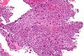
ABC - intermed. mag. (WC/Nephron)
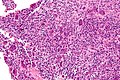
ABC - high mag. (WC/Nephron)
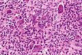
ABC - very high mag. (WC/Nephron)


www:
- ABC - low mag. (webpathology.com).
- ABC - intermed. mag. (webpathology.com).
- ABC - high mag. (webpathology.com).
See also
- Bone.
References
- ↑ 1.0 1.1 URL: http://emedicine.medscape.com/article/1254784-overview. Accessed on: 4 February 2011.
- ↑ van den Berg H, Kroon HM, Slaar A, Hogendoorn P (2008). "Incidence of biopsy-proven bone tumors in children: a report based on the Dutch pathology registration "PALGA"". J Pediatr Orthop 28 (1): 29–35. doi:10.1097/BPO.0b013e3181558cb5. PMID 18157043.
- ↑ Parashari, UC.; Khanduri, S.; Upadhyay, D.; Bhadury, S.; Singhal, S.. "Radiologic and pathologic correlation of aneurysmal bone cysts at unusual sites.". J Cancer Res Ther 8 (1): 103-5. doi:10.4103/0973-1482.95183. PMID 22531523.