Aneurysm
Jump to navigation
Jump to search
An aneurysm is an abnormal blood-filled swelling of an artery or vein, resulting from a localized weakness in the wall of the vessel.
| Aneurysm | |
|---|---|
| Diagnosis in short | |
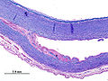
.jpg) Temporal artery aneurysm. H&E stain. | |
| Gross | dilation of blood vessel |
| Site | mostly cerebral, blood vessels |
|
| |
| Clinical history | variable |
| Prognosis | dependent on size, location and subtype |
Aortic aneurysm
Main article: Aortic dissection
- Dilation < 1.5 times [1]
Aortic dissection (WC).

Mycotic pulmonar aneurysm (Yale Rosen).
_Victoria_blue-HE.jpg)
.jpg)
Coronar (heart) aneurysm
Cerebral (intracranial) aneurysm
Clinic
- usu. asymptomatic.
- after rupture: sudden severe headache, nausea, vision impairment, vomiting, loss of consciousness.
Classification
- Size:
- Small < 15 mm, large (15 to 25 mm), giant (25 to 50 mm) and super-giant (> 50 mm).
- Shape:
- Saccular, Fusiform
- Location:

Micro
- Berry aneurysm:
- Fusiform aneurysm:
- often middle segment of basal arteries.
- enlagement: several centimeters.
- marked atreriosclerosis (histiocytes, hemorrhage, calcification, inflammation).
- Infective aneurysm:
- high mortality (30% in bacterial, 90% in fungal).
- seen in 3% of patients with infectious endocarditis.
- history of drug abuse.
- inflammation of the vessel wall (ectasia).
- usu. from the adventitia inwards.
- Presence of microorganisms.
Image
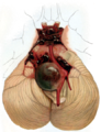
Cerebellar aneurysm (Dr. Bollinger / WC)

See also
References
- ↑ Johnston, KW.; Rutherford, RB.; Tilson, MD.; Shah, DM.; Hollier, L.; Stanley, JC. (Mar 1991). "Suggested standards for reporting on arterial aneurysms. Subcommittee on Reporting Standards for Arterial Aneurysms, Ad Hoc Committee on Reporting Standards, Society for Vascular Surgery and North American Chapter, International Society for Cardiovascular Surgery.". J Vasc Surg 13 (3): 452-8. PMID 1999868.