Alpha-1 antitrypsin deficiency
Jump to navigation
Jump to search
Alpha-1 antitrypsin deficiency, abbreviated A1-AT, is a relatively common genetic condition that causes lung and liver pathology.
It is also known as alpha1-antiprotease inhibitor deficiency.
This article deals with the liver pathology. The lung pathology is panlobular emphysema and covered in the emphysema article.
General
Etiology:
- Genetic defect.
- Prevalence 1 in 2000-5000.[1]
Causes:
- Lung and liver injury.
- Lung -> panlobular emphysema.
Microscopic
Features:
- Pink globules in zone 1 (periportal).
- Globules not seen in children.
- May not be present in late stage (cirrhotic).
- Best seen on PASD stain.
- Can be seen on H&E -- if one looks carefully.
Note:
- The pink globules may be seen in the context of cirrhosis; cases should be confirmation with IHC.
Images
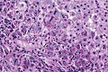
Alpha-1 AT deficiency - PASD. (WC/JMGardner)

www:
Stains
IHC
- A1-AT +ve globules.[3]
See also
References
- ↑ Stoller, JK.; Aboussouan, LS. (Sep 2011). "A Review of Alpha-1 Antitrypsin Deficiency.". Am J Respir Crit Care Med. doi:10.1164/rccm.201108-1428CI. PMID 21960536.
- ↑ 2.0 2.1 Qizilbash, A.; Young-Pong, O. (Jun 1983). "Alpha 1 antitrypsin liver disease differential diagnosis of PAS-positive, diastase-resistant globules in liver cells.". Am J Clin Pathol 79 (6): 697-702. PMID 6189389.
- ↑ Theaker, JM.; Fleming, KA. (Jan 1986). "Alpha-1-antitrypsin and the liver: a routine immunohistological screen.". J Clin Pathol 39 (1): 58-62. PMID 3512609.