Difference between revisions of "Alpha-1 antitrypsin deficiency"
(→Images: added a case) |
(→Images: Added a case with non-specific staining) |
||
| Line 72: | Line 72: | ||
|} | |} | ||
Alpha 1 Antitrypsin deficiency with cirrhosis. Dark inflamed bands bound hepatocyte nodules (Row 1 Left 40X). Steatosis afflicts some hepatocyte areas, within inflamed band lie proliferated bile ductules (Row 1 Right 100X). Reticulin shows regenerative (more than 2 nuclei per cord) nodules (black lines without definite directionality) amid bands (Row 2 Left 100X). Trichrome shows scar (Row 2 Right 100X). Hepatocytes show steatosis, foamy degeneration, and intranuclear inclusions (Row 3 Left 400X). PAS-D shows A1AT cytoplasmic granules; in non-A1AT cirrhosis, these granules would neither be in so many hepatocytes nor, in general, as large (Row 3 Right 400X). | Alpha 1 Antitrypsin deficiency with cirrhosis. Dark inflamed bands bound hepatocyte nodules (Row 1 Left 40X). Steatosis afflicts some hepatocyte areas, within inflamed band lie proliferated bile ductules (Row 1 Right 100X). Reticulin shows regenerative (more than 2 nuclei per cord) nodules (black lines without definite directionality) amid bands (Row 2 Left 100X). Trichrome shows scar (Row 2 Right 100X). Hepatocytes show steatosis, foamy degeneration, and intranuclear inclusions (Row 3 Left 400X). PAS-D shows A1AT cytoplasmic granules; in non-A1AT cirrhosis, these granules would neither be in so many hepatocytes nor, in general, as large (Row 3 Right 400X). | ||
{| | |||
[[File:1 A1AT 2 680x512px.tif| Alpha 1 anti-trypsin (A1AT) granules in cirrhosis, not due to A1AT deficiency; A1AT level was normal.]] | |||
[[File:2 A1AT 2 680x512px.tif| Alpha 1 anti-trypsin (A1AT) granules in cirrhosis, not due to A1AT deficiency; A1AT level was normal.]] | |||
<br> | |||
[[File:3 A1AT 2 680x512px.tif| Alpha 1 anti-trypsin (A1AT) granules in cirrhosis, not due to A1AT deficiency; A1AT level was normal.]] | |||
[[File:4 A1AT 2 680x512px.tif| Alpha 1 anti-trypsin (A1AT) granules in cirrhosis, not due to A1AT deficiency; A1AT level was normal.]] | |||
<br> | |||
[[File:5 A1AT 2 680x512px.tif| Alpha 1 anti-trypsin (A1AT) granules in cirrhosis, not due to A1AT deficiency; A1AT level was normal.]] | |||
[[File:6 A1AT 2 680x512px.tif| Alpha 1 anti-trypsin (A1AT) granules in cirrhosis, not due to A1AT deficiency; A1AT level was normal.]] | |||
|} | |||
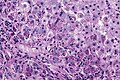
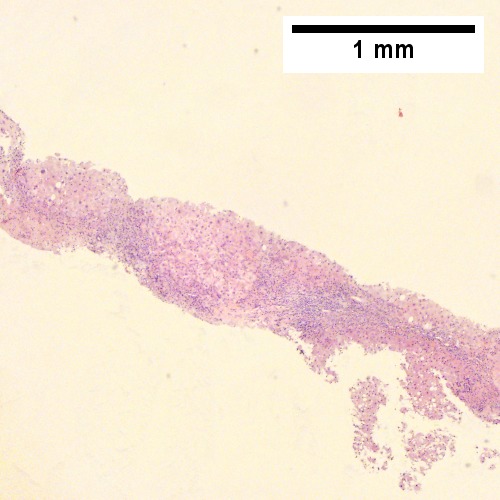
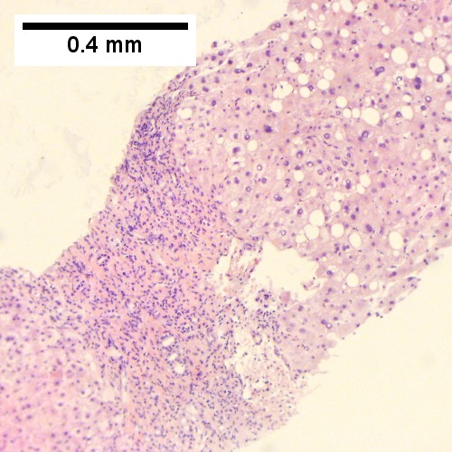
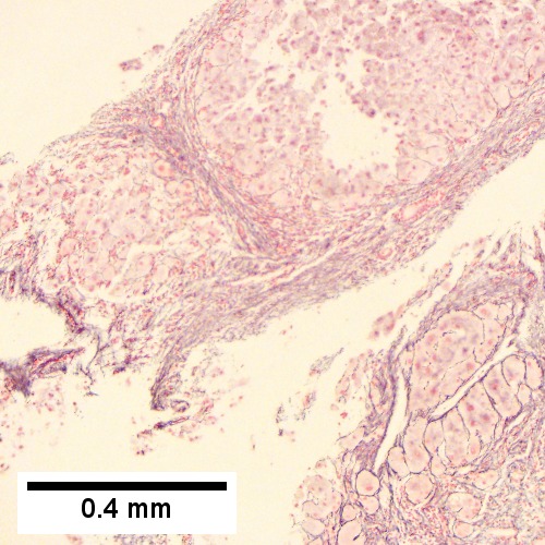
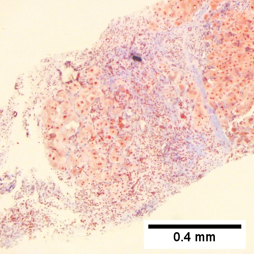
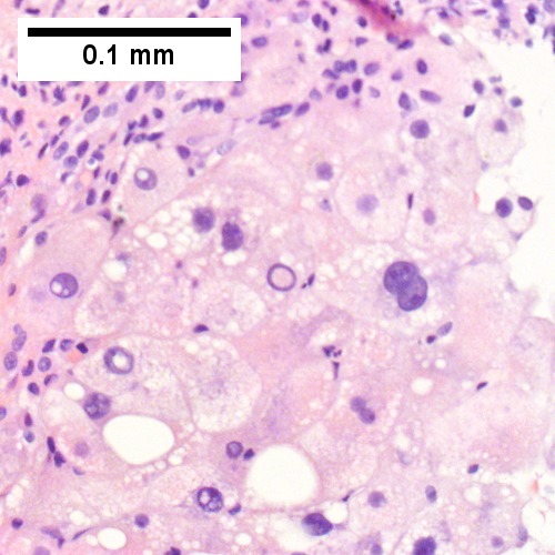
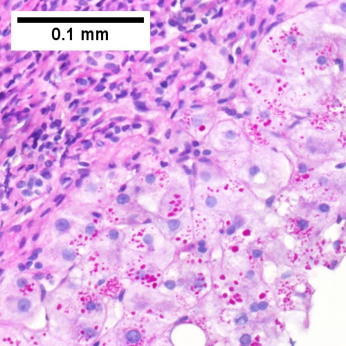
Alpha 1 anti-trypsin (A1AT) granules in cirrhosis, not due to A1AT deficiency; A1AT level was normal. A. Low power shows an inflamed foci, bands, and hepatocyte regions [2x]. B. Trichrome confirms blue fibrosis about hepatocyte isles [4X]. C. Proliferated bile ductules (black arrows) border hepatocytes with glycogenated nuclei (green arrows), consistent with the history of diabetes [200X]. D. Reticulin shows two-cell thick regenerated cords (green arrows) and circles for rosettes (black arrows) [200X]. E. PAS with diastase stain shows occasional small red granules (arrows) and red dust in cytoplasm of hepatocytes near edge of regenerative nodule [400X]. E. A1AT immunostain shows occasional small brown granules (arrows) in cytoplasm of hepatocytes near edge of regenerative nodule [400X]. | |||
www: | www: | ||
Revision as of 16:33, 20 October 2016
| Alpha-1 antitrypsin deficiency | |
|---|---|
| Diagnosis in short | |
 Alpha-1 AT deficiency. PASD stain. | |
|
| |
| Synonyms | alpha1-antiprotease inhibitor deficiency |
|
| |
| LM | +/-pink globules in zone 1 (periportal), +/-fibrosis or cirrhosis |
| Stains | PASD +ve (pink globules in zone 1) - not seen in children |
| IHC | A1-AT +ve globules |
| Site | liver - see medical liver disease, lung - see emphysema |
|
| |
| Prevalence | uncommon (1 in 2000-5000) |
| Radiology | emphysematous changes (chest x-ray) |
Alpha-1 antitrypsin deficiency, abbreviated A1-AT, is a relatively common genetic condition that causes lung and liver pathology.
It is also known as alpha1-antiprotease inhibitor deficiency.
This article deals with the liver pathology. The lung pathology is panlobular emphysema and covered in the emphysema article.
General
Etiology:
- Genetic defect.
- Prevalence 1 in 2000-5000.[1]
Causes:
- Lung and liver injury.
- Lung -> panlobular emphysema.
Microscopic
Features:
- Pink globules in zone 1 (periportal).
- Globules not seen in children.
- May not be present in late stage (cirrhotic).
- Best seen on PASD stain.
- Can be seen on H&E -- if one looks carefully.
Note:
- The pink globules may be seen in the context of cirrhosis; cases should be confirmed with IHC.
Images
Alpha-1 AT deficiency - PASD. (WC/JMGardner)
Alpha 1 Antitrypsin deficiency with cirrhosis. Dark inflamed bands bound hepatocyte nodules (Row 1 Left 40X). Steatosis afflicts some hepatocyte areas, within inflamed band lie proliferated bile ductules (Row 1 Right 100X). Reticulin shows regenerative (more than 2 nuclei per cord) nodules (black lines without definite directionality) amid bands (Row 2 Left 100X). Trichrome shows scar (Row 2 Right 100X). Hepatocytes show steatosis, foamy degeneration, and intranuclear inclusions (Row 3 Left 400X). PAS-D shows A1AT cytoplasmic granules; in non-A1AT cirrhosis, these granules would neither be in so many hepatocytes nor, in general, as large (Row 3 Right 400X).
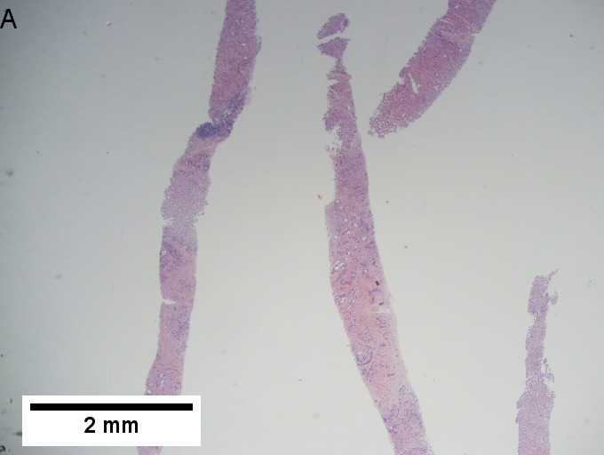
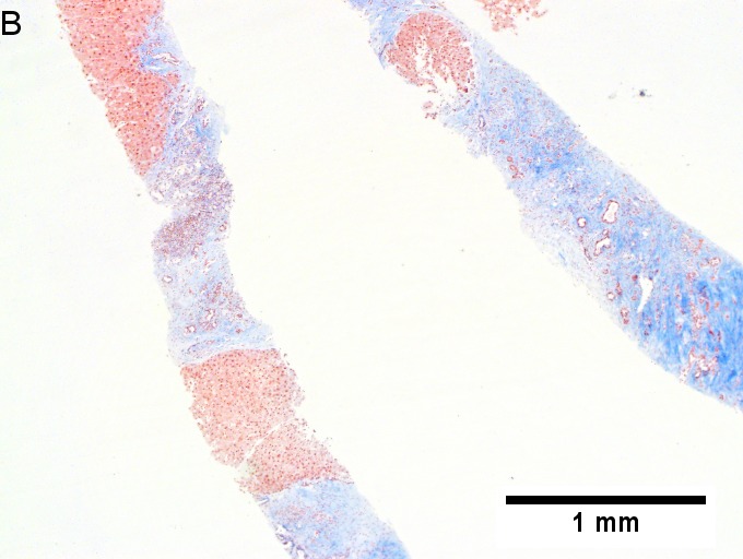
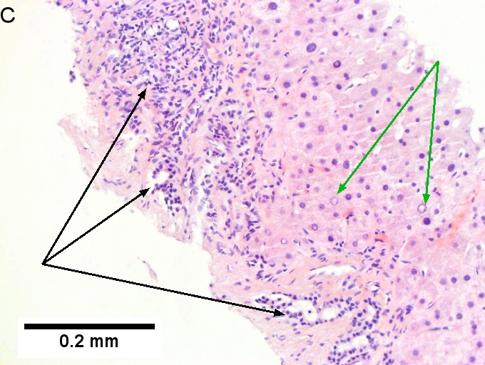
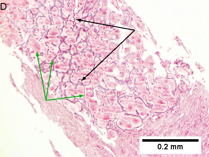
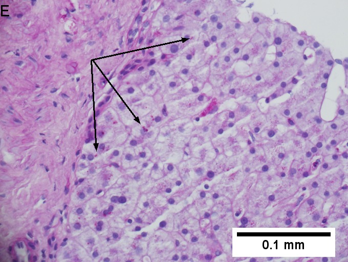
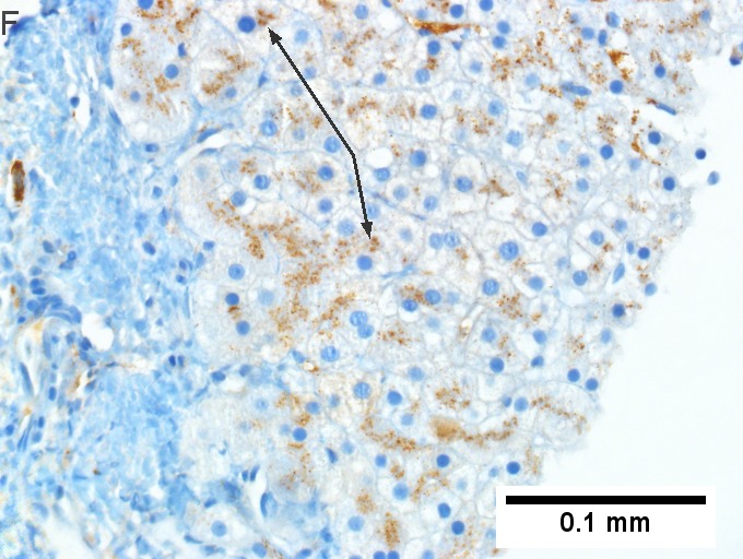
Alpha 1 anti-trypsin (A1AT) granules in cirrhosis, not due to A1AT deficiency; A1AT level was normal. A. Low power shows an inflamed foci, bands, and hepatocyte regions [2x]. B. Trichrome confirms blue fibrosis about hepatocyte isles [4X]. C. Proliferated bile ductules (black arrows) border hepatocytes with glycogenated nuclei (green arrows), consistent with the history of diabetes [200X]. D. Reticulin shows two-cell thick regenerated cords (green arrows) and circles for rosettes (black arrows) [200X]. E. PAS with diastase stain shows occasional small red granules (arrows) and red dust in cytoplasm of hepatocytes near edge of regenerative nodule [400X]. E. A1AT immunostain shows occasional small brown granules (arrows) in cytoplasm of hepatocytes near edge of regenerative nodule [400X].
www:
Stains
IHC
- A1-AT +ve globules.[3]
See also
References
- ↑ Stoller, JK.; Aboussouan, LS. (Sep 2011). "A Review of Alpha-1 Antitrypsin Deficiency.". Am J Respir Crit Care Med. doi:10.1164/rccm.201108-1428CI. PMID 21960536.
- ↑ 2.0 2.1 Qizilbash, A.; Young-Pong, O. (Jun 1983). "Alpha 1 antitrypsin liver disease differential diagnosis of PAS-positive, diastase-resistant globules in liver cells.". Am J Clin Pathol 79 (6): 697-702. PMID 6189389.
- ↑ Theaker, JM.; Fleming, KA. (Jan 1986). "Alpha-1-antitrypsin and the liver: a routine immunohistological screen.". J Clin Pathol 39 (1): 58-62. PMID 3512609.