Adamantinoma
Jump to navigation
Jump to search
The printable version is no longer supported and may have rendering errors. Please update your browser bookmarks and please use the default browser print function instead.
| Adamantinoma | |
|---|---|
| Diagnosis in short | |
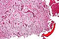
 Adamantinoma. H&E stain. | |
|
| |
| LM | biphasic tumour - epithelial component & fibro-osseous component |
| Subtypes | classic, differentiated |
| LM DDx | vascular tumours (epithelioid hemangioendothelioma), metastatic carcinoma |
| Site | bone - classically tibia, other sites |
|
| |
| Prevalence | uncommon |
| Prognosis | benign +/-locally aggressive |
| Clin. DDx | osteosarcoma |
Adamantinoma is an uncommon benign bone tumour.
It should not be confused with adenomatoid tumour.
General
Features:[1]
- Rare: < 1% of bone tumours.
- Typically 25-35 years old.
- Benign, may be locally aggressive.
Gross
- Classically mid portion of tibia.[2]
- Fibula common.
- Reported in many other sites.
Radiology
- Intracortical, radiolucent.
Microscopic
Features:
- Biphasic tumour:[2]
- Fibro-osseous component.
- Spindle cells.
- Epithelial component.
- Classically nests of basaloid cells.
- Fibro-osseous component.
Note:
- It is described as resembling ameloblastoma,[2] but the resemblance isn't striking.
DDx:[3]
- Vascular tumours (epithelioid hemangioendothelioma).
- Metastatic carcinoma.
Subtypes
Subdivided into:[2]
- Classic.
- Differentiated - less than 20 years old only.
Images
Adamantinoma - intermed. mag. (WC)
www:
- Adamantinoma (southbaypath.org).[4]
- Adamantinoma (nih.gov).[2]
- Adamantinoma (nih.gov).
- Adamantinoma (tumorlibrary.com).
- Adamantinoma (tumorlibrary.com).
- Adamantinoma (tumorlibrary.com).
- Epithelium can be focal - Diagnostico Med Br [1]
![[1]](http://www.diagnostico.med.br/osteopat/54d.JPG)
IHC
Features:[3]
See also
References
- ↑ Humphrey, Peter A; Dehner, Louis P; Pfeifer, John D (2008). The Washington Manual of Surgical Pathology (1st ed.). Lippincott Williams & Wilkins. pp. 650. ISBN 978-0781765275.
- ↑ 2.0 2.1 2.2 2.3 2.4 Jain, D.; Jain, VK.; Vasishta, RK.; Ranjan, P.; Kumar, Y. (2008). "Adamantinoma: a clinicopathological review and update.". Diagn Pathol 3: 8. doi:10.1186/1746-1596-3-8. PMID 18279517.
- ↑ 3.0 3.1 URL: http://www.pathconsultddx.com/pathCon/diagnosis?pii=S1559-8675%2806%2970057-2. Accessed on: 28 April 2011.
- ↑ URL: http://southbaypath.org/CaseImages/sb5260/sb5260.htm. Accessed on: 7 December 2010.
- ↑ URL: http://www.nordiqc.org/Epitopes/Cytokeratins/cytokeratins.htm. Accessed on: 28 April 2011.