Difference between revisions of "Adamantinoma"
Jump to navigation
Jump to search
(more) |
|||
| Line 5: | Line 5: | ||
| Caption = Adamantinoma. [[H&E stain]]. | | Caption = Adamantinoma. [[H&E stain]]. | ||
| Micro = | | Micro = | ||
| Subtypes = | | Subtypes = classic, differentiated | ||
| LMDDx = | | LMDDx = | ||
| Stains = | | Stains = | ||
| Line 34: | Line 34: | ||
*Rare: < 1% of bone tumours. | *Rare: < 1% of bone tumours. | ||
*25-35 years old. | *25-35 years old. | ||
*Benign, may be locally aggressive. | *Benign, may be locally aggressive. | ||
==Gross== | ==Gross== | ||
* | *Classically mid portion of tibia.<ref name=pmid18279517/> | ||
**May be seen in the fibula. | |||
===Radiology=== | ===Radiology=== | ||
| Line 45: | Line 45: | ||
==Microscopic== | ==Microscopic== | ||
Features: | Features: | ||
*Biphasic tumour: | *Biphasic tumour:<ref name=pmid18279517/> | ||
*#Fibrous/spindle cell component. | *#Fibrous/spindle cell component. | ||
*#Epithelial component. | *#Epithelial component. | ||
Note: | |||
*Histologically, it resembles [[ameloblastoma]].<ref name=pmid18279517/> | |||
DDx:<ref name=pathcon_adam>URL: [http://www.pathconsultddx.com/pathCon/diagnosis?pii=S1559-8675%2806%2970057-2 http://www.pathconsultddx.com/pathCon/diagnosis?pii=S1559-8675%2806%2970057-2]. Accessed on: 28 April 2011.</ref> | DDx:<ref name=pathcon_adam>URL: [http://www.pathconsultddx.com/pathCon/diagnosis?pii=S1559-8675%2806%2970057-2 http://www.pathconsultddx.com/pathCon/diagnosis?pii=S1559-8675%2806%2970057-2]. Accessed on: 28 April 2011.</ref> | ||
*Vascular tumours ([[Epithelioid hemangioendothelioma]]). | *Vascular tumours ([[Epithelioid hemangioendothelioma]]). | ||
*[[Metastatic carcinoma]]. | *[[Metastatic carcinoma]]. | ||
===Subtypes=== | |||
Subdivided into:<ref name=pmid18279517/> | |||
*Classic. | |||
*Differentiated - less than 20 years old only. | |||
===Images=== | ===Images=== | ||
Revision as of 22:18, 29 August 2013
| Adamantinoma | |
|---|---|
| Diagnosis in short | |
 Adamantinoma. H&E stain. | |
| Subtypes | classic, differentiated |
| Site | bone - usu. tibia or fibula. |
|
| |
| Prevalence | uncommon |
| Prognosis | benign +/-locally aggressive |
| Clin. DDx | osteosarcoma |
Adamantinoma is an uncommon benign bone tumour. It should not be confused with adenomatoid tumour.
General
Features:[1]
- Rare: < 1% of bone tumours.
- 25-35 years old.
- Benign, may be locally aggressive.
Gross
- Classically mid portion of tibia.[2]
- May be seen in the fibula.
Radiology
- Intracortical, radiolucent.
Microscopic
Features:
- Biphasic tumour:[2]
- Fibrous/spindle cell component.
- Epithelial component.
Note:
- Histologically, it resembles ameloblastoma.[2]
DDx:[3]
- Vascular tumours (Epithelioid hemangioendothelioma).
- Metastatic carcinoma.
Subtypes
Subdivided into:[2]
- Classic.
- Differentiated - less than 20 years old only.
Images
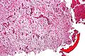
Adamantinoma - intermed. mag. (WC)
www:
IHC
Features:[3]
- CK14 +ve (HMWK).[5]
- CK19 +ve (LMWK).
- CK8/18 -ve (LMWK).
See also
References
- ↑ Humphrey, Peter A; Dehner, Louis P; Pfeifer, John D (2008). The Washington Manual of Surgical Pathology (1st ed.). Lippincott Williams & Wilkins. pp. 650. ISBN 978-0781765275.
- ↑ 2.0 2.1 2.2 2.3 2.4 Jain, D.; Jain, VK.; Vasishta, RK.; Ranjan, P.; Kumar, Y. (2008). "Adamantinoma: a clinicopathological review and update.". Diagn Pathol 3: 8. doi:10.1186/1746-1596-3-8. PMID 18279517.
- ↑ 3.0 3.1 URL: http://www.pathconsultddx.com/pathCon/diagnosis?pii=S1559-8675%2806%2970057-2. Accessed on: 28 April 2011.
- ↑ URL: http://southbaypath.org/CaseImages/sb5260/sb5260.htm. Accessed on: 7 December 2010.
- ↑ URL: http://www.nordiqc.org/Epitopes/Cytokeratins/cytokeratins.htm. Accessed on: 28 April 2011.