Acute myeloid leukemia
Jump to navigation
Jump to search
The printable version is no longer supported and may have rendering errors. Please update your browser bookmarks and please use the default browser print function instead.

Acute myeloid leukemia, abbreviated AML, is a group of malignancies.
Overview
- Group of malignancies.
- Defined by the cytogenetic abnormalities.
No recurrent cytogenetic abnormalities
Acute myeloid leukemia without recurrent cytogenetic abnormalities
General
- Adults.
Exclusions for this diagnosis:
- Prior MDS.
- Down syndrome.
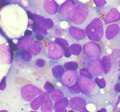
Microscopic
Features:
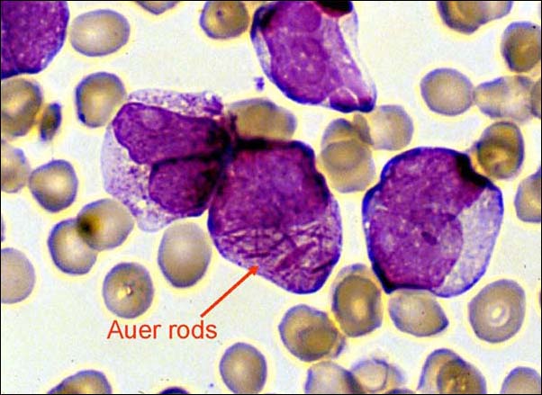
- Auer rods present
- Cytoplasmic granularity.
- Large cells.
Note:
- May be classified by morphology, using the (old) French-American-British (FAB) classification (M0-M7).
Image
Auer rods in an AML. (WC)
www:
Molecular
- Must exclude all the recurrent cytogenetic abnormalities - see below.
AML with recurrent cytogenetic abnormalities
Acute myeloid leukemia with t(8;21)
IHC
- CD34+, CD13+, MPO+ (cytoplasm), CD33+ (weak).
- CD56+, CD117+.
- Usu. assoc. with a bad prognosis.
Flow cytometry
- CD19+, PAX5+, CD79a +/-.
Images:
Molecular
- t(8;21)(q22;q22).[1]
Acute myeloid leukemia with inv(16)
- inv(16)(p13.1q22).[2]
- Associated with myeloid sarcoma.
Microscopic
- Blast count usu. ~20% (low).
- Eosinophilic granules.
- Used to be classified as "M4" with eosinophilia.
IHC
- CD2+ -- common.
Acute myeloid leukemia with t(15;17)
- AKA acute promyelocytic leukemia
- Abbreviated APL.
- t(15;17)(q22;q12).
General
Clinical:
- Associated with DIC.
- Treatment: all-trans retinoic acid (ATRA).
Microscopic
Comes in two flavours.
Microscopic (Hypergranular or typical APL):
- Bean-shaped nucleus or bilobed nucleus.
- Buddles of Auer rods - known as "Faggot cells".
Microscopic (Microgranular or hypogranular APL):
- Bilobed nuclei with nuclear overlap. (???)
- Absence of granules on light microscopy.
Images

Faggot cell in AML-M3. (WC)

www:
IHC
- CD2 +ve, CD34 +ve/-ve, CD56 +ve/-ve.
Flow cytometry
- CD34 -ve, HLA-DR -ve.
- CD33 +ve, CD13 +ve/-ve, CD117 +ve (weak), CD56 +ve/-ve.
Molecular
Variants:
- t(11;17) -- ATRA doesn't work.[6]
- t(17;17) -- ATRA doesn't work.
- t(5;17). (???)
Acute myeloid leukemia with t(9;11)
General
Clinical:
- +/-DIC.
- Usu. children.
Microscopic
- Monoblastic morphology. (???)
- Myelomonocytic morphology. (???)
IHC
- CD33+, CD65+, CD4+, HLA-DR+.
- CD34+. (???)
- CD13+. (???)
Molecular
- t(9;11).
See also
References
- ↑ Berger, R. (1994). "Translocation t(8;21)(q22;q22): cytogenetics and molecular biology.". Nouv Rev Fr Hematol 36 Suppl 1: S67-9. PMID 8177719.
- ↑ Lu, CM.; Murata-Collins, JL.; Wang, E.; Siddiqi, I.; Lawrence, HJ. (Dec 2006). "Concurrent acute myeloid leukemia with inv(16)(p13.1q22) and chronic lymphocytic leukemia: molecular evidence of two separate diseases.". Am J Hematol 81 (12): 963-8. doi:10.1002/ajh.20716. PMID 16917916.
- ↑ Online 'Mendelian Inheritance in Man' (OMIM) 102578
- ↑ Online 'Mendelian Inheritance in Man' (OMIM) 180240
- ↑ URL: http://path.upmc.edu/cases/case457.html. Accessed on: 21 January 2012.
- ↑ Lefkowitch, Jay H. (2006). Anatomic Pathology Board Review (1st ed.). Saunders. pp. 623 Q2. ISBN 978-1416025887.