Chorangioma
Jump to navigation
Jump to search
| Chorangioma | |
|---|---|
| Diagnosis in short | |
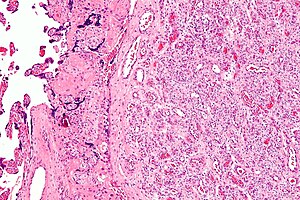
 Chorangioma. H&E stain. | |
|
| |
| LM | abundant capillaries surrounded by stroma |
| LM DDx | chorangiosis, chorangiomatosis |
| Gross | white or red lesion |
| Site | placenta |
|
| |
| Associated Dx | +/-IUGR, +/-fetal hydrops - if large |
| Prevalence | uncommon |
Chorangioma is an uncommon pathology of the placenta that is similar to a hemangioma.
General
- Hamartoma-like growth in the placenta consisting of blood vessels.[1]
Epidemiology:
- Often benign/insignificant; large lesions (>4 cm[1] or >5 cm[2]) or multiple lesions are significant.
- May be association with:
- Fetal maternal haemorrhage.
- Fetal hydrops.
- IUGR.
- Incidence: ~1 in 100 placentas.[1]
Gross
- White lesions.
- Occasionally red lesions.
Microscopic
Features:[1]
- Mass of capillaries - key feature.
- +/-High cellularity.
- +/-Degenerative changes.
Notes:
- Must be differentiated from chorangiomatosis (associated with preeclampsia & IUGR) and chorangiosis (associated with maternal diabetes mellitus).[1]
Images
See also
References
- ↑ 1.0 1.1 1.2 1.3 1.4 Amer HZ, Heller DS (2010). "Chorangioma and related vascular lesions of the placenta--a review". Fetal Pediatr Pathol 29 (4): 199–206. doi:10.3109/15513815.2010.487009. PMID 20594143.
- ↑ Lež C, Fures R, Hrgovic Z, Belina S, Fajdic J, Münstedt K (2010). "Chorangioma placentae". Rare Tumors 2 (4): e67. doi:10.4081/rt.2010.e67. PMC 3019602. PMID 21234259. https://www.ncbi.nlm.nih.gov/pmc/articles/PMC3019602/.

