Difference between revisions of "Adrenal myelolipoma"
Jump to navigation
Jump to search
(+SO) |
|||
| (5 intermediate revisions by 2 users not shown) | |||
| Line 1: | Line 1: | ||
{{ Infobox diagnosis | {{ Infobox diagnosis | ||
| Name = {{PAGENAME}} | | Name = {{PAGENAME}} | ||
| Image = Myelolipoma_histology_HE.jpg | | Image = Myelolipoma_histology_HE.jpg | ||
| Width = | | Width = | ||
| Caption = Myelolipoma. [[H&E stain]]. | | Caption = Myelolipoma. [[H&E stain]]. | ||
| Line 61: | Line 61: | ||
<gallery> | <gallery> | ||
Image:Myelolipoma_histology_HE.jpg | Myelolipoma (WC/Mattopaedia) | Image:Myelolipoma_histology_HE.jpg | Myelolipoma (WC/Mattopaedia) | ||
Image:Adrenal Myelolipoma LP CTR.jpg|Adrenal myelolipoma - low power. (SKB) | |||
Image:Adrenal Myelolipoma MP CTR (2).jpg|Adrenal myelolipoma - medium power - Adrenal gland to the right. (SKB) | |||
Image:Adrenal Myelolipoma MP CTR.jpg|Adrenal Myelolipoma - medium power - Adrenal gland to the left. (SKB) | |||
Image:Adrenal Myelolipoma HP CTR.jpg|Adrenal myelolipoma - high power. (SKB) | |||
Image:Adrenal Myelolipoma HP2 CTR.jpg|Adrenal myelolipoma - high power. (SKB) | |||
</gallery> | </gallery> | ||
| Line 66: | Line 71: | ||
*[http://www.ncbi.nlm.nih.gov/pmc/articles/PMC3162227/figure/F3/ Myelolipoma (nih.gov)].<ref name=pmid21927708/> | *[http://www.ncbi.nlm.nih.gov/pmc/articles/PMC3162227/figure/F3/ Myelolipoma (nih.gov)].<ref name=pmid21927708/> | ||
*[http://path.upmc.edu/cases/case165.html Adrenal myelolipoma (upmc.edu)]. | *[http://path.upmc.edu/cases/case165.html Adrenal myelolipoma (upmc.edu)]. | ||
==Sign out== | |||
<pre> | |||
A. Right Adrenal Gland, Adrenalectomy: | |||
- Adrenal myelolipoma. | |||
</pre> | |||
===Microscopic=== | |||
The sections shows a lesion with bland adipocytes and hematopoietic elements (erythroid, myeloid, megakaryocytic) that is surrounded by a thin rim of adrenal cortex and a partial fibrous pseudocapsule. Significant cellular atypia is absent. The lesion appears to be clear of the inked margin. | |||
==See also== | ==See also== | ||
Latest revision as of 21:07, 16 May 2024
| Adrenal myelolipoma | |
|---|---|
| Diagnosis in short | |
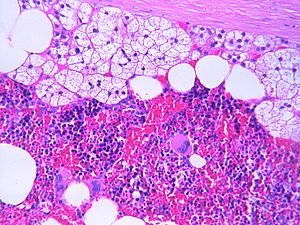
 Myelolipoma. H&E stain. | |
|
| |
| LM | adipose tissue, hematopoietic elements from all three lineages (erythroid, myeloid, megakaryocytic), +/-calcification |
| LM DDx | angiomyolipoma of the kidney, lipoma, liposarcoma, teratoma |
| Site | adrenal gland |
|
| |
| Prevalence | rare |
| Prognosis | benign |
| Clin. DDx | other adrenal tumours (e.g. adrenal cortical carcinoma), retroperitoneal tumours |
| Treatment | excision if large |
Adrenal myelolipoma is a benign tumour of the adrenal gland.
Myelolipoma redirects here.
General
- Benign and rare.
- Typically asymptomatic and hormonally inactive.[1]
- Symptoms: back or abdominal pain.
- Diagnosis - usu. by abdominal CT.
Treatment:
- Watchful waiting if small (<=7 cm) and asymptomatic.[1]
Microscopic
Features:[2]
- Adipose tissue.
- Hematopoietic elements from all three lineages:
- Erythroid.
- Myeloid.
- Megakaryocytic.
- +/-Calcification.[1]
DDx:[3]
- Angiomyolipoma of the kidney.
- Lipoma.
- Liposarcoma.
- Teratoma.
Images
www:
Sign out
A. Right Adrenal Gland, Adrenalectomy:
	- Adrenal myelolipoma.
Microscopic
The sections shows a lesion with bland adipocytes and hematopoietic elements (erythroid, myeloid, megakaryocytic) that is surrounded by a thin rim of adrenal cortex and a partial fibrous pseudocapsule. Significant cellular atypia is absent. The lesion appears to be clear of the inked margin.
See also
References
- ↑ 1.0 1.1 1.2 Daneshmand, S.; Quek, ML. (2006). "Adrenal myelolipoma: diagnosis and management.". Urol J 3 (2): 71-4. PMID 17590837.
- ↑ 2.0 2.1 Cha, JS.; Shin, YS.; Kim, MK.; Kim, HJ. (Aug 2011). "Myelolipomas of both adrenal glands.". Korean J Urol 52 (8): 582-5. doi:10.4111/kju.2011.52.8.582. PMC 3162227. PMID 21927708. https://www.ncbi.nlm.nih.gov/pmc/articles/PMC3162227/.
- ↑ Lam, KY.; Lo, CY. (Sep 2001). "Adrenal lipomatous tumours: a 30 year clinicopathological experience at a single institution.". J Clin Pathol 54 (9): 707-12. PMID 11533079.


