Squamous cell carcinoma of the esophagus
| Squamous cell carcinoma of the esophagus | |
|---|---|
| Diagnosis in short | |
 Esophageal squamous cell carcinoma. H&E stain. | |
|
| |
| LM | atypical squamous cells invading into the lamina propria |
| LM DDx | squamous dysplasia of the esophagus |
| IHC | p63 +'ve, CK5/6 +'ve |
| Site | esophagus - usually proximal or mid portion |
|
| |
| Associated Dx | squamous dysplasia of the esophagus, alcohol, smoking |
| Symptoms | dysphagia of solid and liquids |
| Prevalence | uncommon |
| Prognosis | poor |
| Clin. DDx | benign esophageal stricture |
| Treatment | usu. surgery - if feasible, chemotherapy, radiation |
Squamous cell carcinoma of the esophagus is a relatively uncommon form of esophageal cancer.
It is also known as esophageal squamous cell carcinoma, abbreviated esophageal SCC.
Squamous cell carcinoma, also squamous carcinoma, is discussed in general terms in the squamous cell carcinoma article.
General
- Like squamous cell carcinoma elsewhere.
Risk factors:[1]
- Alcohol consumption.
- Tobacco use.
- Food with nitrosamines.
- Burning-hot beverages.
- Disputed.[2]
Note:
- Reflux is not a risk factor for esophageal SCC.
- It is a risk factor indirectly for esophageal adenocarcinoma.
Clinical:
- Dysphagia of solid and liquids.
- Weight loss.
- Multimodal treatment - surgery, chemotherapy and radiation.[3]
Gross
- Mass in the esophagus - classically proximal or mid portion.
Microscopic
Features - atypical squamous cells with invasion through the basement membrane:
- Cytology:
- Nucleus - typical central.
- +/-Mitoses.
- Cytoplasm - "dense-appearing", typically eosinophilic (may be intensely eosinophilic).
- Nucleus - typical central.
- +/-Squamous whorls.
Note:
- Just to make things confusing, the Staging of early SCC differs from that of early adenocarcinoma!
DDx:
- Reactive changes.
- Squamous dysplasia of the esophagus.
- Adenocarcinoma of the esophagus.
- Metastatic carcinoma.
Images
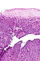
Esophageal SCC - intermed. mag.
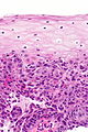
Esophageal SCC - high mag.
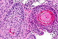
Esophageal SCC - very high mag.


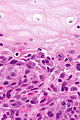
Esophageal SCC - high mag.
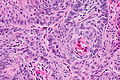
Esophageal SCC - high mag.
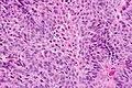
Esophageal SCC - high mag.
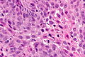
Esophageal SCC - very high mag.


www:
Sign out
ESOPHAGUS, BIOPSY:
- INVASIVE SQUAMOUS CELL CARCINOMA, KERATINIZING, MODERATELY DIFFERENTIATED.

COMMENT:
Benign squamous epithelium at least partially overlies the invasive squamous
cell carcinoma; this may mask the true extent of the lesion on endoscopy.
Micro
The sections show a squamous mucosa with focal moderate atypia of the squamous cells, keratinization and easily identified mitotic figures. The atypical cells are partially covered by benign squamous cells, and there is a very sharp transition between the cells with atypia and those without. The atypical squamous cells extend into the subepithelial tissue in irregularly shaped nests and cords. A small amount of benign muscle is present.
See also
References
- ↑ Lefkowitch, Jay H. (2006). Anatomic Pathology Board Review (1st ed.). Saunders. pp. 104 Q1. ISBN 978-1416025887.
- ↑ Zamora-Ros, R.; Luján-Barroso, L.; Bueno-de-Mesquita, HB.; Dik, VK.; Boeing, H.; Steffen, A.; Tjønneland, A.; Olsen, A. et al. (Feb 2014). "Tea and coffee consumption and risk of esophageal cancer: the European Prospective Investigation into Cancer and Nutrition (EPIC) study.". Int J Cancer. doi:10.1002/ijc.28789. PMID 24535727.
- ↑ Bass, GA.; Furlong, H.; O'Sullivan, KE.; Hennessy, TP.; Walsh, TN. (Jan 2014). "Chemoradiotherapy, with adjuvant surgery for local control, confers a durable survival advantage in adenocarcinoma and squamous cell carcinoma of the oesophagus.". Eur J Cancer. doi:10.1016/j.ejca.2013.12.022. PMID 24480403.
- ↑ Terada, T. (2013). "A clinicopathologic study of esophageal 860 benign and malignant lesions in 910 cases of consecutive esophageal biopsies.". Int J Clin Exp Pathol 6 (2): 191-8. PMID 23330004.