Disordered proliferative endometrium
| Disordered proliferative endometrium | |
|---|---|
| Diagnosis in short | |
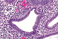
 Disordered proliferative endometrium. H&E stain. | |
|
| |
| LM | proliferative endometrial glands (pseudostratified nuclei + mitoses) with focally abnormal glands (glands >2x normal size; irregular shape -- typically with inflection points; >4 glands involved (dilated)), +/-stromal condensation, gland-to-stromal ratio normal, not within an endometrial polyp |
| LM DDx | proliferative phase endometrium,simple endometrial hyperplasia, benign endometrial polyp |
| Site | endometrium |
|
| |
| Prognosis | benign |
| Treatment | followup - re-biopsy |
Disordered proliferative endometrium, abbreviated DPE, is an abnormal endometrial finding with some features of simple endometrial hyperplasia.
General
- Association: anovulation.
- Benign - can be grouped with normal.[1]
Treatment:
Image:
Microscopic
Features:[4]
- Proliferative type endometrium with:
- Cystic dilation of glands focally that do not have (glandular) secretions - key feature.
- Glands >2x normal size - usually 3-4x normal.
- Irregular shape, e.g. gland contour has inflection points.
- Greater than four glands involved (dilated).
- Cystic dilation of glands focally that do not have (glandular) secretions - key feature.
- +/-Stromal condensation -- balls of stromal tissue, aka "blue balls" (due to breakdown of endometrium).
Notes:
- Dilated glands often have tubal metaplasia.[citation needed]
- Eosinophilic syncytial metaplasia - common.
- Features: abundant eosinophilic cytoplasm, mild nuclear atypia +/-loss of nuclear stratification, no mitoses).
DDx:
- Proliferative phase endometrium.
- Glands: straight, tubular, tall pseudostratified columnar cells, mitotic figures, no vacuolation, no mucus secretion, abundant mitoses.
- Stroma: cellular, stroma (spindle cells), mitoses.
- Simple endometrial hyperplasia without atypia - architectural atypia diffuse.
- Benign endometrial polyp - may have gland dilation.
- Anovulatory endometrium - some consider this a synonym, see relation to disordered proliferative endometrium.
Images
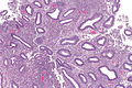
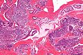
DPE - low mag.
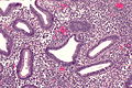
DPE - intermed. mag.
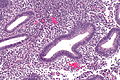
DPE - intermed. mag.
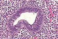
DPE - high mag.
DPE - high mag.


Endometrial stromal condensation - high mag. (WC/Nephron)

www:
Sign out
ENDOMETRIUM, BIOPSY:
- DISORDERED PROLIFERATIVE ENDOMETRIUM.
With endocervix
ENDOMETRIUM, BIOPSY:
- DISORDERED PROLIFERATIVE ENDOMETRIUM.
- BENIGN ENDOCERVICAL MUCOSA.
Waffle a bit
ENDOMETRIUM, BIOPSY:
- COMPATIBLE WITH DISORDERED PROLIFERATIVE ENDOMETRIUM (FRAGMENTS OF PROLIFERATIVE
  ENDOMETRIUM WITH EVIDENCE OF SHEDDING AND VERY RARE GLAND DILATION).
- VERY SCANT STRIPPED ENDOCERVICAL EPITHELIUM WITHOUT APPARENT PATHOLOGY.
- NEGATIVE FOR ENDOMETRIAL HYPERPLASIA.
- NEGATIVE FOR MALIGNANCY.
ENDOMETRIUM, CURETTAGE:
- PROLIFERATIVE ENDOMETRIUM, FOCALLY WITH GLAND DILATION AND SMALL BLOOD
  VESSELS, SEE COMMENT.
- NEGATIVE FOR HYPERPLASIA AND NEGATIVE FOR MALIGNANCY.

COMMENT:
A fibrotic stroma is not present. The findings may represent a remnant of the previously
excised endometrial polyp or disordered proliferative endometrium. Follow-up is suggested.
Micro
The sections show a well-sampled endometrium. Mitotic figures are identified within the glands and stroma. Irregular, moderately enlarged glands are seen (only) in one of several fragments; most of the endometrial glands are round, regular and small.
No stromal condensation is apparent. No secretions are in the glands.
There are no back-to-back glands. No nuclear atypia is apparent. No thick-walled blood vessels are apparent.
See also
References
- ↑ Sherman, ME.; Ronnett, BM.; Ioffe, OB.; Richesson, DA.; Rush, BB.; Glass, AG.; Chatterjee, N.; Duggan, MA. et al. (Jul 2008). "Reproducibility of biopsy diagnoses of endometrial hyperplasia: evidence supporting a simplified classification.". Int J Gynecol Pathol 27 (3): 318-25. doi:10.1097/PGP.0b013e3181659167. PMID 18580308.
- ↑ McCluggage, WG. (Aug 2006). "My approach to the interpretation of endometrial biopsies and curettings.". J Clin Pathol 59 (8): 801-12. doi:10.1136/jcp.2005.029702. PMID 16873562.
- ↑ 3.0 3.1 Ely, JW.; Kennedy, CM.; Clark, EC.; Bowdler, NC.. "Abnormal uterine bleeding: a management algorithm.". J Am Board Fam Med 19 (6): 590-602. PMID 17090792. http://www.jabfm.org/content/19/6/590.full.
- ↑ Cotran, Ramzi S.; Kumar, Vinay; Fausto, Nelson; Nelso Fausto; Robbins, Stanley L.; Abbas, Abul K. (2005). Robbins and Cotran pathologic basis of disease (7th ed.). St. Louis, Mo: Elsevier Saunders. pp. 1080 and 1082. ISBN 0-7216-0187-1.
- ↑ URL: http://www.glowm.com/index.html?p=glowm.cml/section_view&articleid=235. Accessed on: 11 December 2012.