Difference between revisions of "Zoon vulvitis"
Jump to navigation
Jump to search
| (14 intermediate revisions by 2 users not shown) | |||
| Line 1: | Line 1: | ||
{{ Infobox diagnosis | |||
| Name = {{PAGENAME}} | |||
| Image = Plasmacytosis mucosae -- high mag.jpg | |||
| Width = | |||
| Caption = Zoon vulvitis. [[H&E stain]]. | |||
| Synonyms = [[plasmacytosis mucosae]] | |||
| Micro = squamous epithelium with: spongiosis, lack of ''stratum granulosum'' and ''stratum corneum'' layers, "lozenge"-shaped (diamond-like shape); abundant plasma cells - esp. superficial dermis; +/-hemosiderin deposits | |||
| Subtypes = | |||
| LMDDx = [[syphilis]], fungal infection | |||
| Stains = PASF -ve | |||
| IHC = | |||
| EM = | |||
| Molecular = | |||
| IF = | |||
| Gross = | |||
| Grossing = | |||
| Site = [[vulva]] | |||
| Assdx = | |||
| Syndromes = | |||
| Clinicalhx = | |||
| Signs = | |||
| Symptoms = pruritis, pain | |||
| Prevalence = | |||
| Bloodwork = | |||
| Rads = | |||
| Endoscopy = | |||
| Prognosis = | |||
| Other = | |||
| ClinDDx = | |||
| Tx = | |||
}} | |||
'''Zoon vulvitis''', also '''vulvitis circumscripta plasmacellularis'''<ref>{{Cite journal | last1 = dos Reis | first1 = HL. | last2 = de Vargas | first2 = PR. | last3 = Lucas | first3 = E. | last4 = Camporez | first4 = T. | last5 = Ferreira | first5 = Dde C. | title = Zoon vulvitis as a differential diagnosis in an HIV-infected patient: a short report. | journal = J Int Assoc Provid AIDS Care | volume = 12 | issue = 3 | pages = 159-61 | month = | year = | doi = 10.1177/2325957412467694 | PMID = 23449712 }}</ref> and '''plasma cell vulvitis''', is a rare pathology of the [[vulva]]. | '''Zoon vulvitis''', also '''vulvitis circumscripta plasmacellularis'''<ref>{{Cite journal | last1 = dos Reis | first1 = HL. | last2 = de Vargas | first2 = PR. | last3 = Lucas | first3 = E. | last4 = Camporez | first4 = T. | last5 = Ferreira | first5 = Dde C. | title = Zoon vulvitis as a differential diagnosis in an HIV-infected patient: a short report. | journal = J Int Assoc Provid AIDS Care | volume = 12 | issue = 3 | pages = 159-61 | month = | year = | doi = 10.1177/2325957412467694 | PMID = 23449712 }}</ref> and '''plasma cell vulvitis''', is a rare pathology of the [[vulva]]. | ||
It is also known as ''[[plasmacytosis mucosae]]'',<ref name=Ref_GP11>{{Ref GP|11}}</ref> which may be used to refer to ''[[Zoon balanitis]]''. | |||
==General== | ==General== | ||
| Line 8: | Line 41: | ||
*Pruritis. | *Pruritis. | ||
*Pain. | *Pain. | ||
*Orange colouration.<ref>URL: [http://www.dermnetnz.org/site-age-specific/plasma-cell.html http://www.dermnetnz.org/site-age-specific/plasma-cell.html]. Accessed on: October 29, 2014.</ref> | |||
==Gross== | ==Gross== | ||
| Line 20: | Line 54: | ||
**"Lozenge"-shaped (diamond-like shape). | **"Lozenge"-shaped (diamond-like shape). | ||
*Abundant plasma cells - esp. superficial dermis. | *Abundant plasma cells - esp. superficial dermis. | ||
**Keep in mind that plasma cells are a common component of mucosal infiltrates, much more so than for skin. | |||
**A plasma cell component has to be relatively prominent to be relevant in this location (the same can be said for eosinophils). | |||
*Hemosiderin deposits.<ref name=pmid6833541>{{Cite journal | last1 = Davis | first1 = J. | last2 = Shapiro | first2 = L. | last3 = Baral | first3 = J. | title = Vulvitis circumscripta plasmacellularis. | journal = J Am Acad Dermatol | volume = 8 | issue = 3 | pages = 413-6 | month = Mar | year = 1983 | doi = | PMID = 6833541 }}</ref> | *Hemosiderin deposits.<ref name=pmid6833541>{{Cite journal | last1 = Davis | first1 = J. | last2 = Shapiro | first2 = L. | last3 = Baral | first3 = J. | title = Vulvitis circumscripta plasmacellularis. | journal = J Am Acad Dermatol | volume = 8 | issue = 3 | pages = 413-6 | month = Mar | year = 1983 | doi = | PMID = 6833541 }}</ref> | ||
DDx: | DDx: | ||
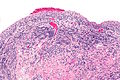
*[[Syphilis]]. | *[[Syphilis]] - Spirochete immunostain (in red) is helpful. | ||
*Lichenoid drug reaction. | *Spongiotic vulvitis. | ||
**Might look very similar. | |||
**Usually wont show iron deposition. | |||
**Clinical correlation important. | |||
*Superficial fungal vulvitis (usually Candida). | |||
*Lichenoid vulvitis (lichen planus/lichenoid drug reaction). | |||
**Both can be very plasmocellular on mucosal surfaces. | |||
**Zoon vulvitis won't show prominent lichenoid destruction of the epithelium. | |||
====Images==== | ====Images==== | ||
<gallery> | |||
Image: Plasmacytosis mucosae -- intermed mag.jpg | PM - intermed. mag. | |||
Image: Plasmacytosis mucosae -- high mag.jpg | PM - high mag. | |||
Image: Plasmacytosis mucosae - alt -- high mag.jpg | PM - high mag. | |||
Image: Plasmacytosis mucosae -- very high mag.jpg | PM - very high mag. | |||
</gallery> | |||
www: | |||
*[http://www.surgicalpathologyatlas.com/glfusion/mediagallery/media.php?s=20080802172103984 Vulvitis chronica plasmacellularis (surgicalpathologyatlas.com)]. | *[http://www.surgicalpathologyatlas.com/glfusion/mediagallery/media.php?s=20080802172103984 Vulvitis chronica plasmacellularis (surgicalpathologyatlas.com)]. | ||
*[http://www.ijdvl.com/viewimage.asp?img=ijdvl_2012_78_2_230_93664_f2.jpg Zoon vulvitis (ijdvl.com)].<ref name=pmid22421677/> | *[http://www.ijdvl.com/viewimage.asp?img=ijdvl_2012_78_2_230_93664_f2.jpg Zoon vulvitis (ijdvl.com)].<ref name=pmid22421677/> | ||
| Line 46: | Line 96: | ||
A CD138 immunostain demonstrates clusters of plasma cells. A p16 immunostain | A CD138 immunostain demonstrates clusters of plasma cells. A p16 immunostain | ||
is negative. Ki-67 positive cells are basal. No microorganisms are apparent | is negative. Ki-67 positive cells are basal. No microorganisms are apparent | ||
with a PASF stain. | with a PASF stain. Perls stain positive iron is admixed with the inflammatory infiltrate. | ||
</pre> | </pre> | ||
Latest revision as of 03:43, 14 November 2014
| Zoon vulvitis | |
|---|---|
| Diagnosis in short | |
 Zoon vulvitis. H&E stain. | |
|
| |
| Synonyms | plasmacytosis mucosae |
|
| |
| LM | squamous epithelium with: spongiosis, lack of stratum granulosum and stratum corneum layers, "lozenge"-shaped (diamond-like shape); abundant plasma cells - esp. superficial dermis; +/-hemosiderin deposits |
| LM DDx | syphilis, fungal infection |
| Stains | PASF -ve |
| Site | vulva |
|
| |
| Symptoms | pruritis, pain |
Zoon vulvitis, also vulvitis circumscripta plasmacellularis[1] and plasma cell vulvitis, is a rare pathology of the vulva.
It is also known as plasmacytosis mucosae,[2] which may be used to refer to Zoon balanitis.
General
- Rare ~ 50 reported cases in English literature as of 2012.[3]
- Analogous to Zoon's balanitis in males.[4]
Clinical:[3]
- Pruritis.
- Pain.
- Orange colouration.[5]
Gross
Features:[3]
- Erythematous macules or papules.
Microscopic
Features:
- Squamous epithelium with:[2]
- Spongiosis.
- Lack of stratum granulosum and stratum corneum layers.
- "Lozenge"-shaped (diamond-like shape).
- Abundant plasma cells - esp. superficial dermis.
- Keep in mind that plasma cells are a common component of mucosal infiltrates, much more so than for skin.
- A plasma cell component has to be relatively prominent to be relevant in this location (the same can be said for eosinophils).
- Hemosiderin deposits.[6]
DDx:
- Syphilis - Spirochete immunostain (in red) is helpful.
- Spongiotic vulvitis.
- Might look very similar.
- Usually wont show iron deposition.
- Clinical correlation important.
- Superficial fungal vulvitis (usually Candida).
- Lichenoid vulvitis (lichen planus/lichenoid drug reaction).
- Both can be very plasmocellular on mucosal surfaces.
- Zoon vulvitis won't show prominent lichenoid destruction of the epithelium.
Images
PM - intermed. mag.
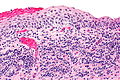
PM - high mag.
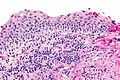
PM - high mag.
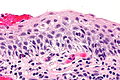
PM - very high mag.


www:
IHC
- CD138 -- to highlight plasma cells.
Sign out
LESION, VULVA, BIOPSY:
- SQUAMOUS MUCOSA WITHOUT A CORNIFIED ZONE, WITH MARKED CHRONIC INFLAMMATION
  WITH PROMINENT PLASMA CELLS, SEE COMMENT.
- NEGATIVE FOR DYSPLASIA AND NEGATIVE FOR MALIGNANCY.

COMMENT:
The loss of the cornified zone and prominent plasma cells is consistent
with plasma cell vulvitis (Zoon vulvitis). Clinical correlation is suggested.

A CD138 immunostain demonstrates clusters of plasma cells. A p16 immunostain
is negative. Ki-67 positive cells are basal. No microorganisms are apparent
with a PASF stain.  Perls stain positive iron is admixed with the inflammatory infiltrate.
See also
References
- ↑ dos Reis, HL.; de Vargas, PR.; Lucas, E.; Camporez, T.; Ferreira, Dde C.. "Zoon vulvitis as a differential diagnosis in an HIV-infected patient: a short report.". J Int Assoc Provid AIDS Care 12 (3): 159-61. doi:10.1177/2325957412467694. PMID 23449712.
- ↑ 2.0 2.1 Nucci, Marisa R.; Oliva, Esther (2009). Gynecologic Pathology: A Volume in Foundations in Diagnostic Pathology Series (1st ed.). Churchill Livingstone. pp. 11. ISBN 978-0443069208.
- ↑ 3.0 3.1 3.2 3.3 Çelik, A.; Haliloglu, B.; Tanriöver, Y.; Ilter, E.; Gündüz, T.; Ulu, I.; Midi, A.; Özekici, Ü.. "Plasma cell vulvitis: a vulvar itching dilemma.". Indian J Dermatol Venereol Leprol 78 (2): 230. doi:10.4103/0378-6323.93664. PMID 22421677.
- ↑ Yoganathan, S.; Bohl, TG.; Mason, G. (Dec 1994). "Plasma cell balanitis and vulvitis (of Zoon). A study of 10 cases.". J Reprod Med 39 (12): 939-44. PMID 7884748.
- ↑ URL: http://www.dermnetnz.org/site-age-specific/plasma-cell.html. Accessed on: October 29, 2014.
- ↑ Davis, J.; Shapiro, L.; Baral, J. (Mar 1983). "Vulvitis circumscripta plasmacellularis.". J Am Acad Dermatol 8 (3): 413-6. PMID 6833541.