Difference between revisions of "Esophageal adenocarcinoma"
Jump to navigation
Jump to search
| (23 intermediate revisions by the same user not shown) | |||
| Line 1: | Line 1: | ||
{{ Infobox diagnosis | |||
| Name = {{PAGENAME}} | |||
| Image = Esophageal_adenocarcinoma_-_high_mag.jpg | |||
| Width = | |||
| Caption = Esophageal adenocarcinoma. [[H&E stain]]. | |||
| Synonyms = | |||
| Micro = "invading" cell clusters or glands (gland cribriforming (more than rare) or desmoplasia or submucosa invasion); nuclear atypia of malignancy (variation of size, shape, staining), +/-mitoses | |||
| Subtypes = | |||
| LMDDx = [[high-grade columnar dysplasia of the esophagus]], [[squamous cell carcinoma of the esophagus]],[[gastric adenocarcinoma]] | |||
| Stains = | |||
| IHC = CK7 +ve, CK20 -ve, p63 -ve | |||
| EM = | |||
| Molecular = | |||
| IF = | |||
| Gross = | |||
| Grossing = | |||
| Site = [[esophagus]] - classically distal esophagus | |||
| Assdx = [[Barrett's esophagus]], [[high-grade columnar dysplasia of the esophagus]] | |||
| Syndromes = | |||
| Clinicalhx = | |||
| Signs = | |||
| Symptoms = dysphagia | |||
| Prevalence = uncommon | |||
| Bloodwork = | |||
| Rads = | |||
| Endoscopy = | |||
| Prognosis = often poor - as typically high stage at diagnosis | |||
| Other = | |||
| ClinDDx = | |||
| Tx = [[EMR]] (early), surgery - esophagectomy | |||
}} | |||
'''Esophageal adenocarcinoma''', also '''adenocarcinoma of the esophagus''', is a common malignant epithelial-derived tumour of the distal esophagus, that classically arises in the context of [[Barrett's esophagus]]. | '''Esophageal adenocarcinoma''', also '''adenocarcinoma of the esophagus''', is a common malignant epithelial-derived tumour of the distal esophagus, that classically arises in the context of [[Barrett's esophagus]]. | ||
| Line 5: | Line 36: | ||
*May be difficult to distinguish from adenocarcinoma of the stomach. | *May be difficult to distinguish from adenocarcinoma of the stomach. | ||
**By convention (in the ''[[CAP checklist]]'') gastroesophageal junction carcinomas are staged as esophageal carcinomas.<ref>URL: [http://www.cap.org/apps/docs/committees/cancer/cancer_protocols/2011/Esophagus_11protocol.pdf http://www.cap.org/apps/docs/committees/cancer/cancer_protocols/2011/Esophagus_11protocol.pdf]. Accessed on: 6 April 2012.</ref> | **By convention (in the ''[[CAP checklist]]'') gastroesophageal junction carcinomas are staged as esophageal carcinomas.<ref>URL: [http://www.cap.org/apps/docs/committees/cancer/cancer_protocols/2011/Esophagus_11protocol.pdf http://www.cap.org/apps/docs/committees/cancer/cancer_protocols/2011/Esophagus_11protocol.pdf]. Accessed on: 6 April 2012.</ref> | ||
Symptoms: | |||
*Dysphagia - difficulty swallowing. | |||
Associations: | |||
*[[Barrett's esophagus]]. | |||
**[[Gastroesophageal reflux disease]]. | |||
*Hiatal hernia.<ref name=pmid24495377>{{cite journal |author=Jiang X, Tseng CC, Bernstein L, Wu AH |title=Family history of cancer and gastroesophageal disorders and risk of esophageal and gastric adenocarcinomas: a case-control study |journal=BMC Cancer |volume=14 |issue= |pages=60 |year=2014 |pmid=24495377 |pmc=3915076 |doi=10.1186/1471-2407-14-60 |url=}}</ref> | |||
*Family history - [[prostate cancer]].<ref name=pmid24495377/> | |||
===Treatment=== | ===Treatment=== | ||
*Adenocarcinoma in situ (AIS) - may be treated with endoscopic mucosal resection & follow-up.<ref name=pmid19306943>{{Cite journal | last1 = Sampliner | first1 = RE. | title = Endoscopic therapy for Barrett's esophagus. | journal = Clin Gastroenterol Hepatol | volume = 7 | issue = 7 | pages = 716-20 | month = Jul | year = 2009 | doi = 10.1016/j.cgh.2009.03.011 | PMID = 19306943 }}</ref> | *Adenocarcinoma in situ (AIS) - may be treated with [[endoscopic mucosal resection]] & follow-up.<ref name=pmid19306943>{{Cite journal | last1 = Sampliner | first1 = RE. | title = Endoscopic therapy for Barrett's esophagus. | journal = Clin Gastroenterol Hepatol | volume = 7 | issue = 7 | pages = 716-20 | month = Jul | year = 2009 | doi = 10.1016/j.cgh.2009.03.011 | PMID = 19306943 }}</ref> | ||
*Surgery - esophagectomy. | *Surgery - esophagectomy. | ||
| Line 18: | Line 58: | ||
Features: | Features: | ||
*Adenocarcinoma: | *Adenocarcinoma: | ||
** | **"Invading" cell clusters or glands. | ||
***Cribriforming (more than rare) or desmoplasia or "deep" invasion (into submucosa). | |||
**Nuclear atypia of malignancy: | **Nuclear atypia of malignancy: | ||
***Size variation. | ***Size variation. | ||
***Shape variation. | ***Shape variation. | ||
***Staining variation. | ***Staining variation. | ||
**Mitoses common. | **+/-Mitoses (common). | ||
DDx: | DDx: | ||
*[[Columnar dysplasia of the esophagus|High-grade columnar dysplasia of the esophagus]] | *[[Columnar dysplasia of the esophagus|High-grade columnar dysplasia of the esophagus]] - see table in ''[[esophagus]]'' article | ||
*[[Squamous cell carcinoma of the esophagus]]. | *[[Squamous cell carcinoma of the esophagus]]. | ||
*[[Gastric adenocarcinoma]]. | *[[Gastric adenocarcinoma]]. | ||
| Line 33: | Line 74: | ||
<gallery> | <gallery> | ||
Image:Esophageal_adenocarcinoma_-_very_low_mag.jpg |Esophageal adenocarcinoma - very low mag. (WC) | Image:Esophageal_adenocarcinoma_-_very_low_mag.jpg |Esophageal adenocarcinoma - very low mag. (WC) | ||
Image:Esophageal_adenocarcinoma_-_low_mag.jpg |Esophageal adenocarcinoma - low mag. (WC) | |||
Image:Esophageal_adenocarcinoma_-_intermed_mag.jpg |Esophageal adenocarcinoma - intermed. mag. (WC) | Image:Esophageal_adenocarcinoma_-_intermed_mag.jpg |Esophageal adenocarcinoma - intermed. mag. (WC) | ||
Image:Esophageal_adenocarcinoma_-_high_mag.jpg |Esophageal adenocarcinoma - high mag. (WC) | |||
</gallery> | </gallery> | ||
| Line 43: | Line 86: | ||
====Staging==== | ====Staging==== | ||
Early esophageal adenocarcinoma has its own staging system:<ref>{{Cite journal | last1 = Pech | first1 = O. | last2 = May | first2 = A. | last3 = Rabenstein | first3 = T. | last4 = Ell | first4 = C. | title = Endoscopic resection of early oesophageal cancer. | journal = Gut | volume = 56 | issue = 11 | pages = 1625-34 | month = Nov | year = 2007 | doi = 10.1136/gut.2006.112110 | PMID = 17938435 | PMC = 2095648 }}</ref><ref>{{Cite journal | last1 = Thosani | first1 = N. | last2 = Singh | first2 = H. | last3 = Kapadia | first3 = A. | last4 = Ochi | first4 = N. | last5 = Lee | first5 = JH. | last6 = Ajani | first6 = J. | last7 = Swisher | first7 = SG. | last8 = Hofstetter | first8 = WL. | last9 = Guha | first9 = S. | title = Diagnostic accuracy of EUS in differentiating mucosal versus submucosal invasion of superficial esophageal cancers: a systematic review and meta-analysis. | journal = Gastrointest Endosc | volume = | issue = | pages = | month = Nov | year = 2011 | doi = 10.1016/j.gie.2011.09.016 | PMID = 22115605 | URL = http://www.sciencedirect.com/science/article/pii/S0016510711022048 }}</ref> | Early esophageal adenocarcinoma has its own staging system:<ref name=pmid17938435 >{{Cite journal | last1 = Pech | first1 = O. | last2 = May | first2 = A. | last3 = Rabenstein | first3 = T. | last4 = Ell | first4 = C. | title = Endoscopic resection of early oesophageal cancer. | journal = Gut | volume = 56 | issue = 11 | pages = 1625-34 | month = Nov | year = 2007 | doi = 10.1136/gut.2006.112110 | PMID = 17938435 | PMC = 2095648 }}</ref><ref name=pmid22115605>{{Cite journal | last1 = Thosani | first1 = N. | last2 = Singh | first2 = H. | last3 = Kapadia | first3 = A. | last4 = Ochi | first4 = N. | last5 = Lee | first5 = JH. | last6 = Ajani | first6 = J. | last7 = Swisher | first7 = SG. | last8 = Hofstetter | first8 = WL. | last9 = Guha | first9 = S. | title = Diagnostic accuracy of EUS in differentiating mucosal versus submucosal invasion of superficial esophageal cancers: a systematic review and meta-analysis. | journal = Gastrointest Endosc | volume = | issue = | pages = | month = Nov | year = 2011 | doi = 10.1016/j.gie.2011.09.016 | PMID = 22115605 | URL = http://www.sciencedirect.com/science/article/pii/S0016510711022048 }}</ref> | ||
*M1 = lamina propria. | *M1 = lamina propria. | ||
*M2 = superficial muscularis mucosae. | *M2 = superficial muscularis mucosae. | ||
*M3 = submucosa. | *M3 = submucosa. | ||
*M4 = muscularis propria. | *M4 = muscularis propria. | ||
<!-- | |||
Comment: | |||
*Different staging systems exist; confusingly, they use the same terms (M1, M2, M3).<ref name=pmid15557945>{{Cite journal | last1 = Buskens | first1 = CJ. | last2 = Westerterp | first2 = M. | last3 = Lagarde | first3 = SM. | last4 = Bergman | first4 = JJ. | last5 = ten Kate | first5 = FJ. | last6 = van Lanschot | first6 = JJ. | title = Prediction of appropriateness of local endoscopic treatment for high-grade dysplasia and early adenocarcinoma by EUS and histopathologic features. | journal = Gastrointest Endosc | volume = 60 | issue = 5 | pages = 703-10 | month = Nov | year = 2004 | doi = | PMID = 15557945 }}</ref> --> | |||
==IHC== | ==IHC== | ||
*CK7 +ve. | Features:<ref name=pmid14631371>{{cite journal |authors=Driessen A, Nafteux P, Lerut T, Van Raemdonck D, De Leyn P, Filez L, Penninckx F, Geboes K, Ectors N |title=Identical cytokeratin expression pattern CK7+/CK20- in esophageal and cardiac cancer: etiopathological and clinical implications |journal=Mod Pathol |volume=17 |issue=1 |pages=49–55 |date=January 2004 |pmid=14631371 |doi=10.1038/modpathol.3800011 |url=}}</ref> | ||
*CK20 +ve. | *CK7 +ve (54 of 66 cases). | ||
*CK20 -ve (14 of 66 cases). | |||
Note: | |||
*CK7 +ve and CK20 -ve is seen in ~67% cases (44/66).<ref name=pmid14631371/> | |||
To rule-out SCC: | To rule-out SCC: | ||
*p63 -ve. | *p63 -ve. | ||
*HWMK -ve. | *HWMK -ve. | ||
Others:<ref name=pmid17650224>{{Cite journal | last1 = Liu | first1 = YS. | last2 = Yu | first2 = CH. | last3 = Li | first3 = L. | last4 = Zhang | first4 = BF. | last5 = Fang | first5 = J. | last6 = Zhou | first6 = Q. | last7 = Hu | first7 = Y. | last8 = Li | first8 = YM. | last9 = Jun Gao | first9 = H. | title = Expression of p53, p16 and cyclooxygenase-2 in esophageal cancer with tissue microarray. | journal = J Dig Dis | volume = 8 | issue = 3 | pages = 133-8 | month = Aug | year = 2007 | doi = 10.1111/j.1443-9573.2007.00299.x | PMID = 17650224 }}</ref> | |||
*p53 +ve. | |||
*COX-2 +ve. | |||
*p16 +ve - but not that useful as it's frequently positive in the precursors.<ref name=pmid15617840>{{Cite journal | last1 = Hardie | first1 = LJ. | last2 = Darnton | first2 = SJ. | last3 = Wallis | first3 = YL. | last4 = Chauhan | first4 = A. | last5 = Hainaut | first5 = P. | last6 = Wild | first6 = CP. | last7 = Casson | first7 = AG. | title = p16 expression in Barrett's esophagus and esophageal adenocarcinoma: association with genetic and epigenetic alterations. | journal = Cancer Lett | volume = 217 | issue = 2 | pages = 221-30 | month = Jan | year = 2005 | doi = 10.1016/j.canlet.2004.06.025 | PMID = 15617840 }}</ref> | |||
==Staging== | |||
*The number of lymph nodes is important for [[staging]], as a small number may lead to stage migration (Will Rogers phenomenon). | |||
*There is no established standard for esophageal cancer (as per UICC/AJCC staging - based on Li ''et al.''<ref name=pmid23124992>{{Cite journal | last1 = Li | first1 = Q. | last2 = Wu | first2 = SG. | last3 = Gao | first3 = JM. | last4 = Xu | first4 = JJ. | last5 = Hu | first5 = LY. | last6 = Xu | first6 = T. | title = Impact of esophageal cancer staging on overall survival and disease-free survival based on the 2010 AJCC classification by lymph nodes. | journal = J Radiat Res | volume = 54 | issue = 2 | pages = 307-14 | month = Mar | year = 2013 | doi = 10.1093/jrr/rrs096 | PMID = 23124992 }}</ref>), several studies give different numbers (18 lymph nodes Greenstein ''et al.'',<ref name=pmid18224663>{{Cite journal | last1 = Greenstein | first1 = AJ. | last2 = Litle | first2 = VR. | last3 = Swanson | first3 = SJ. | last4 = Divino | first4 = CM. | last5 = Packer | first5 = S. | last6 = Wisnivesky | first6 = JP. | title = Effect of the number of lymph nodes sampled on postoperative survival of lymph node-negative esophageal cancer. | journal = Cancer | volume = 112 | issue = 6 | pages = 1239-46 | month = Mar | year = 2008 | doi = 10.1002/cncr.23309 | PMID = 18224663 }}</ref> 23 lymph nodes Peyre ''et al.''<ref name=pmid18936567>{{Cite journal | last1 = Peyre | first1 = CG. | last2 = Hagen | first2 = JA. | last3 = DeMeester | first3 = SR. | last4 = Altorki | first4 = NK. | last5 = Ancona | first5 = E. | last6 = Griffin | first6 = SM. | last7 = Hölscher | first7 = A. | last8 = Lerut | first8 = T. | last9 = Law | first9 = S. | title = The number of lymph nodes removed predicts survival in esophageal cancer: an international study on the impact of extent of surgical resection. | journal = Ann Surg | volume = 248 | issue = 4 | pages = 549-56 | month = Oct | year = 2008 | doi = 10.1097/SLA.0b013e318188c474 | PMID = 18936567 }}</ref>). | |||
==Sign out== | ==Sign out== | ||
===Biopsy=== | |||
<pre> | |||
Esophagus, Biopsy: | |||
- INVASIVE ADENOCARCINOMA, poorly differentiated. | |||
Comment: | |||
Pending IHC (CK7, CK20) and HER2 testing; results of these will be reported in an addendum. | |||
</pre> | |||
===Resection=== | |||
<pre> | <pre> | ||
GASTROESOPHAGEAL JUNCTION, RESECTION: | GASTROESOPHAGEAL JUNCTION, RESECTION: | ||
| Line 76: | Line 145: | ||
==See also== | ==See also== | ||
*[[Esophagus]]. | *[[Esophagus]]. | ||
*[[Esophageal cancer]]. | |||
*[[Adenocarcinoma]]. | |||
*[[Siewert classification]]. | |||
==References== | ==References== | ||
Latest revision as of 16:29, 30 January 2024
| Esophageal adenocarcinoma | |
|---|---|
| Diagnosis in short | |
 Esophageal adenocarcinoma. H&E stain. | |
|
| |
| LM | "invading" cell clusters or glands (gland cribriforming (more than rare) or desmoplasia or submucosa invasion); nuclear atypia of malignancy (variation of size, shape, staining), +/-mitoses |
| LM DDx | high-grade columnar dysplasia of the esophagus, squamous cell carcinoma of the esophagus,gastric adenocarcinoma |
| IHC | CK7 +ve, CK20 -ve, p63 -ve |
| Site | esophagus - classically distal esophagus |
|
| |
| Associated Dx | Barrett's esophagus, high-grade columnar dysplasia of the esophagus |
| Symptoms | dysphagia |
| Prevalence | uncommon |
| Prognosis | often poor - as typically high stage at diagnosis |
| Treatment | EMR (early), surgery - esophagectomy |
Esophageal adenocarcinoma, also adenocarcinoma of the esophagus, is a common malignant epithelial-derived tumour of the distal esophagus, that classically arises in the context of Barrett's esophagus.
General
- Often a prognosis poor - as diagnosed in a late stage.
- May be difficult to distinguish from adenocarcinoma of the stomach.
- By convention (in the CAP checklist) gastroesophageal junction carcinomas are staged as esophageal carcinomas.[1]
Symptoms:
- Dysphagia - difficulty swallowing.
Associations:
- Barrett's esophagus.
- Hiatal hernia.[2]
- Family history - prostate cancer.[2]
Treatment
- Adenocarcinoma in situ (AIS) - may be treated with endoscopic mucosal resection & follow-up.[3]
- Surgery - esophagectomy.
Esophagus versus stomach
The convention is it's esophageal if both of the following are true:[4]
- Epicenter of tumour is in the esophagus.
- Barrett's mucosa is present.
Microscopic
Features:
- Adenocarcinoma:
- "Invading" cell clusters or glands.
- Cribriforming (more than rare) or desmoplasia or "deep" invasion (into submucosa).
- Nuclear atypia of malignancy:
- Size variation.
- Shape variation.
- Staining variation.
- +/-Mitoses (common).
- "Invading" cell clusters or glands.
DDx:
- High-grade columnar dysplasia of the esophagus - see table in esophagus article
- Squamous cell carcinoma of the esophagus.
- Gastric adenocarcinoma.
Images

Esophageal adenocarcinoma - very low mag. (WC)
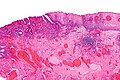
Esophageal adenocarcinoma - low mag. (WC)
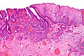
Esophageal adenocarcinoma - intermed. mag. (WC)
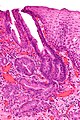
Esophageal adenocarcinoma - high mag. (WC)


Grading
Graded like other adenocarcinoma:[4]
- >95 % of tumour in glandular arrangement = well-differentiated.
- 95-50% of tumour in glandular arrangement= moderately-differentiated.
- <50% of tumour in glandular arrangment = poorly-differentiated.
Staging
Early esophageal adenocarcinoma has its own staging system:[5][6]
- M1 = lamina propria.
- M2 = superficial muscularis mucosae.
- M3 = submucosa.
- M4 = muscularis propria.
IHC
Features:[7]
- CK7 +ve (54 of 66 cases).
- CK20 -ve (14 of 66 cases).
Note:
- CK7 +ve and CK20 -ve is seen in ~67% cases (44/66).[7]
To rule-out SCC:
- p63 -ve.
- HWMK -ve.
Others:[8]
- p53 +ve.
- COX-2 +ve.
- p16 +ve - but not that useful as it's frequently positive in the precursors.[9]
Staging
- The number of lymph nodes is important for staging, as a small number may lead to stage migration (Will Rogers phenomenon).
- There is no established standard for esophageal cancer (as per UICC/AJCC staging - based on Li et al.[10]), several studies give different numbers (18 lymph nodes Greenstein et al.,[11] 23 lymph nodes Peyre et al.[12]).
Sign out
Biopsy
Esophagus, Biopsy:
- INVASIVE ADENOCARCINOMA, poorly differentiated.

Comment:
Pending IHC (CK7, CK20) and HER2 testing; results of these will be reported in an addendum.
Resection
GASTROESOPHAGEAL JUNCTION, RESECTION:
- INTRAMUCOSAL ADENOCARCINOMA, pT1a, pNx, SEE TUMOUR SUMMARY.
-- MUCOSAL MARGIN HAS CAUTERIZED DYSPLASTIC EPITHELIUM, SEE COMMENT.
-- DEEP MARGIN NEGATIVE FOR MALIGNANCY.

COMMENT:
The cauterized dysplastic epithelium cannot be further interpreted.
Malignant-appearing glands are within 1 mm of the cauterized epithelium.
Close follow-up and re-biopsy (or endoscopic re-resection if clinically
indicated) is recommended.

This case was partially reviewed internally. There is agreement on the presence of
intramucosal adenocarcinoma and dysplastic epithelium at the mucosal margin.
See also
References
- ↑ URL: http://www.cap.org/apps/docs/committees/cancer/cancer_protocols/2011/Esophagus_11protocol.pdf. Accessed on: 6 April 2012.
- ↑ 2.0 2.1 Jiang X, Tseng CC, Bernstein L, Wu AH (2014). "Family history of cancer and gastroesophageal disorders and risk of esophageal and gastric adenocarcinomas: a case-control study". BMC Cancer 14: 60. doi:10.1186/1471-2407-14-60. PMC 3915076. PMID 24495377. https://www.ncbi.nlm.nih.gov/pmc/articles/PMC3915076/.
- ↑ Sampliner, RE. (Jul 2009). "Endoscopic therapy for Barrett's esophagus.". Clin Gastroenterol Hepatol 7 (7): 716-20. doi:10.1016/j.cgh.2009.03.011. PMID 19306943.
- ↑ 4.0 4.1 Humphrey, Peter A; Dehner, Louis P; Pfeifer, John D (2008). The Washington Manual of Surgical Pathology (1st ed.). Lippincott Williams & Wilkins. pp. 168. ISBN 978-0781765275.
- ↑ Pech, O.; May, A.; Rabenstein, T.; Ell, C. (Nov 2007). "Endoscopic resection of early oesophageal cancer.". Gut 56 (11): 1625-34. doi:10.1136/gut.2006.112110. PMC 2095648. PMID 17938435. https://www.ncbi.nlm.nih.gov/pmc/articles/PMC2095648/.
- ↑ Thosani, N.; Singh, H.; Kapadia, A.; Ochi, N.; Lee, JH.; Ajani, J.; Swisher, SG.; Hofstetter, WL. et al. (Nov 2011). "Diagnostic accuracy of EUS in differentiating mucosal versus submucosal invasion of superficial esophageal cancers: a systematic review and meta-analysis.". Gastrointest Endosc. doi:10.1016/j.gie.2011.09.016. PMID 22115605.
- ↑ 7.0 7.1 Driessen A, Nafteux P, Lerut T, Van Raemdonck D, De Leyn P, Filez L, Penninckx F, Geboes K, Ectors N (January 2004). "Identical cytokeratin expression pattern CK7+/CK20- in esophageal and cardiac cancer: etiopathological and clinical implications". Mod Pathol 17 (1): 49–55. doi:10.1038/modpathol.3800011. PMID 14631371.
- ↑ Liu, YS.; Yu, CH.; Li, L.; Zhang, BF.; Fang, J.; Zhou, Q.; Hu, Y.; Li, YM. et al. (Aug 2007). "Expression of p53, p16 and cyclooxygenase-2 in esophageal cancer with tissue microarray.". J Dig Dis 8 (3): 133-8. doi:10.1111/j.1443-9573.2007.00299.x. PMID 17650224.
- ↑ Hardie, LJ.; Darnton, SJ.; Wallis, YL.; Chauhan, A.; Hainaut, P.; Wild, CP.; Casson, AG. (Jan 2005). "p16 expression in Barrett's esophagus and esophageal adenocarcinoma: association with genetic and epigenetic alterations.". Cancer Lett 217 (2): 221-30. doi:10.1016/j.canlet.2004.06.025. PMID 15617840.
- ↑ Li, Q.; Wu, SG.; Gao, JM.; Xu, JJ.; Hu, LY.; Xu, T. (Mar 2013). "Impact of esophageal cancer staging on overall survival and disease-free survival based on the 2010 AJCC classification by lymph nodes.". J Radiat Res 54 (2): 307-14. doi:10.1093/jrr/rrs096. PMID 23124992.
- ↑ Greenstein, AJ.; Litle, VR.; Swanson, SJ.; Divino, CM.; Packer, S.; Wisnivesky, JP. (Mar 2008). "Effect of the number of lymph nodes sampled on postoperative survival of lymph node-negative esophageal cancer.". Cancer 112 (6): 1239-46. doi:10.1002/cncr.23309. PMID 18224663.
- ↑ Peyre, CG.; Hagen, JA.; DeMeester, SR.; Altorki, NK.; Ancona, E.; Griffin, SM.; Hölscher, A.; Lerut, T. et al. (Oct 2008). "The number of lymph nodes removed predicts survival in esophageal cancer: an international study on the impact of extent of surgical resection.". Ann Surg 248 (4): 549-56. doi:10.1097/SLA.0b013e318188c474. PMID 18936567.