Difference between revisions of "Embryonal carcinoma"
Jump to navigation
Jump to search
(create) |
(split-out to own article) |
||
| Line 1: | Line 1: | ||
# | {{ Infobox diagnosis | ||
| Name = {{PAGENAME}} | |||
| Image = Embryonal_carcinoma_-_very_high_mag_-_cropped.jpg | |||
| Width = | |||
| Caption = Embryonal carcinoma. [[H&E stain]]. | |||
| Micro = vesicular nuclei, nuclear overlap, necrosis (common), mitoses, variable architecture (tubulopapillary, glandular, solid, embryoid bodies) | |||
| Subtypes = | |||
| LMDDx = [[seminoma]], [[mixed germ cell tumour]], other carcinomas | |||
| Stains = | |||
| IHC = CD30 +ve, AE1/AE3 +ve | |||
| EM = | |||
| Molecular = | |||
| IF = | |||
| Gross = | |||
| Grossing = | |||
| Site = [[testis]], [[ovary]] | |||
| Assdx = | |||
| Syndromes = | |||
| Signs = testicular mass, pelvic mass | |||
| Symptoms = | |||
| Prevalence = | |||
| Bloodwork = | |||
| Rads = | |||
| Endoscopy = | |||
| Prognosis = | |||
| Other = | |||
| ClinDDx = | |||
}} | |||
'''Embryonal carcinoma''' is a type of [[germ cell tumour]]. It is commonly as a component of [[mixed germ cell tumour]]s. | |||
==General== | |||
*Affects young adults. | |||
**May be seen in women. | |||
==Microscopic== | |||
Features:<ref name=Ref_GUP549>{{Ref GUP|549}}</ref> | |||
#Nucleoli - '''key feature'''. | |||
#Vesicular nuclei (clear, empty appearing nuclei) - '''key feature'''. | |||
#Nuclei overlap. | |||
#[[Necrosis]] - common. | |||
#*Not commonly present in seminoma. | |||
#Indistinct cell borders | |||
#Mitoses - common. | |||
#Variable architecture: | |||
#*Tubulopapillary. | |||
#*Glandular. | |||
#*Solid. | |||
#*Embryoid bodies - ball of cells in surrounded by empty space on three sides. | |||
Notes: | |||
*Cytoplasmic staining variable (eosinophilic to basophilic). | |||
DDx: | |||
*[[Yolk sac tumour]]. | |||
===Images=== | |||
<gallery> | |||
Image:Embryonal_carcinoma_-_very_high_mag_-_cropped.jpg | Embryonal carcinoma - very high mag. - cropped (WC/Nephron) | |||
Image:Embryonal_carcinoma_-_high_mag.jpg | Embryonal carcinoma - high mag. (WC/Nephron) | |||
</gallery> | |||
<gallery> | |||
Image:Embryonal_carcinoma_high_mag.jpg | Embryonal carcinoma - high mag. (WC/Nephron) | |||
Image:Embryonal_carcinoma_intermed_mag.jpg | Embryonal carcinoma - intermed. mag. (WC/Nephron) | |||
Image:Embryonal_carcinoma_low_mag.jpg | Embryonal carcinoma - low mag. (WC/Nephron) | |||
</gallery> | |||
==IHC== | |||
*AE1/AE3 +ve. | |||
*CD30 +ve. | |||
==See also== | |||
*[[Germ cell tumours]]. | |||
*[[Ovarian tumours]]. | |||
*[[Testis]]. | |||
==References== | |||
{{Reflist|1}} | |||
[[Category:Diagnosis]] | [[Category:Diagnosis]] | ||
Revision as of 07:24, 3 July 2013
| Embryonal carcinoma | |
|---|---|
| Diagnosis in short | |
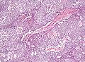
 Embryonal carcinoma. H&E stain. | |
|
| |
| LM | vesicular nuclei, nuclear overlap, necrosis (common), mitoses, variable architecture (tubulopapillary, glandular, solid, embryoid bodies) |
| LM DDx | seminoma, mixed germ cell tumour, other carcinomas |
| IHC | CD30 +ve, AE1/AE3 +ve |
| Site | testis, ovary |
|
| |
| Signs | testicular mass, pelvic mass |
Embryonal carcinoma is a type of germ cell tumour. It is commonly as a component of mixed germ cell tumours.
General
- Affects young adults.
- May be seen in women.
Microscopic
Features:[1]
- Nucleoli - key feature.
- Vesicular nuclei (clear, empty appearing nuclei) - key feature.
- Nuclei overlap.
- Necrosis - common.
- Not commonly present in seminoma.
- Indistinct cell borders
- Mitoses - common.
- Variable architecture:
- Tubulopapillary.
- Glandular.
- Solid.
- Embryoid bodies - ball of cells in surrounded by empty space on three sides.
Notes:
- Cytoplasmic staining variable (eosinophilic to basophilic).
DDx:
Images
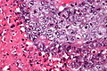
Embryonal carcinoma - very high mag. - cropped (WC/Nephron)
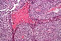
Embryonal carcinoma - high mag. (WC/Nephron)

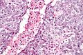
Embryonal carcinoma - high mag. (WC/Nephron)
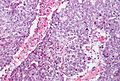
Embryonal carcinoma - intermed. mag. (WC/Nephron)
Embryonal carcinoma - low mag. (WC/Nephron)


IHC
- AE1/AE3 +ve.
- CD30 +ve.
See also
References
- ↑ Zhou, Ming; Magi-Galluzzi, Cristina (2006). Genitourinary Pathology: A Volume in Foundations in Diagnostic Pathology Series (1st ed.). Churchill Livingstone. pp. 549. ISBN 978-0443066771.