Difference between revisions of "Cardiomyopathy"
Jump to navigation
Jump to search
(→Hypertrophic cardiomyopathy: more) |
|||
| (19 intermediate revisions by the same user not shown) | |||
| Line 10: | Line 10: | ||
==Dilated cardiomyopathy== | ==Dilated cardiomyopathy== | ||
*Abbreviated ''DCM''. | |||
===General=== | ===General=== | ||
*Most common of the cardiomyopathies. | *Most common of the cardiomyopathies. | ||
| Line 17: | Line 17: | ||
*Myocarditis - leading cause, usually viral.<ref name=pmid19017683>{{cite journal |author=Luk A, Ahn E, Soor GS, Butany J |title=Dilated cardiomyopathy: a review |journal=J. Clin. Pathol. |volume=62 |issue=3 |pages=219–25 |year=2009 |month=March |pmid=19017683 |doi=10.1136/jcp.2008.060731 |url=}}</ref> | *Myocarditis - leading cause, usually viral.<ref name=pmid19017683>{{cite journal |author=Luk A, Ahn E, Soor GS, Butany J |title=Dilated cardiomyopathy: a review |journal=J. Clin. Pathol. |volume=62 |issue=3 |pages=219–25 |year=2009 |month=March |pmid=19017683 |doi=10.1136/jcp.2008.060731 |url=}}</ref> | ||
*Familial ~ 30% - can be AD with variable penetrance, AR, X-linked. | *Familial ~ 30% - can be AD with variable penetrance, AR, X-linked. | ||
*In the [[forensic pathology|forensic]] context, usually caused by alcoholism.<ref name=Ref_HoFP43>{{Ref HoFP|43}}</ref> | *In the [[forensic pathology|forensic]] context, usually caused by [[alcoholism]].<ref name=Ref_HoFP43>{{Ref HoFP|43}}</ref> | ||
===Microscopic=== | ===Microscopic=== | ||
| Line 25: | Line 25: | ||
DDx: | DDx: | ||
*Mitochondrial myopathy. | *[[Mitochondrial myopathy]]. | ||
**Perinuclear clearing on light microscopy due to abundant mitochondria. | **Perinuclear clearing on light microscopy due to abundant mitochondria. | ||
**Atypical mitochondria on [[electron microscopy]]. | **Atypical mitochondria on [[electron microscopy]]. | ||
*Muscular dystrophy. | *[[Muscular dystrophy]]. | ||
*[[Storage disease]]. | *[[Storage disease]]. | ||
| Line 43: | Line 43: | ||
===General=== | ===General=== | ||
*Genetic. | *Genetic. | ||
*Classic cause of sudden death in young athletes.<ref name=pmid17330410>{{cite journal |author=Gojanovic B, Feihl F, Gremion G, Waeber B |title=[Sudden death in young athletes] |language=German |journal=Praxis (Bern 1994) |volume=96 |issue=6 |pages=189-98 |year=2007 |month=February |pmid=17330410 |doi= |url=}}</ref> | |||
===Gross=== | ===Gross=== | ||
| Line 60: | Line 61: | ||
Images: | Images: | ||
*[http:// | *[http://medicinembbs.blogspot.com/2011/03/hypertrophic-obstructive-cardiomyopathy.html HCM (blogspot.com)]. | ||
*[http:// | *[http://medcell.med.yale.edu/histology/muscle/hypertrophic_cardiomyopathy.php HCM (yale.edu)]. | ||
===Variants=== | ===Variants=== | ||
====Hypertrophic obstructive cardiomyopathy==== | ====Hypertrophic obstructive cardiomyopathy==== | ||
*Abbreviated ''HOCM''. | |||
*Considered to be a variant of HCM. | *Considered to be a variant of HCM. | ||
*Historically known as ''idiopathic hypertrophic subaortic stenosis'' (IHSS). | *Historically known as ''idiopathic hypertrophic subaortic stenosis'' (IHSS). | ||
| Line 78: | Line 80: | ||
===Etiology=== | ===Etiology=== | ||
Multiple causes - an incomplete list:<ref name=Ref_HoFP44-5>{{Ref HoFP|44-5}}</ref> | Multiple causes - an incomplete list:<ref name=Ref_HoFP44-5>{{Ref HoFP|44-5}}</ref> | ||
*[[Hemochromatosis]]. | *[[Hemochromatosis]] - rare.<ref name=pmid7446557>{{Cite journal | last1 = Cutler | first1 = DJ. | last2 = Isner | first2 = JM. | last3 = Bracey | first3 = AW. | last4 = Hufnagel | first4 = CA. | last5 = Conrad | first5 = PW. | last6 = Roberts | first6 = WC. | last7 = Kerwin | first7 = DM. | last8 = Weintraub | first8 = AM. | title = Hemochromatosis heart disease: an unemphasized cause of potentially reversible restrictive cardiomyopathy. | journal = Am J Med | volume = 69 | issue = 6 | pages = 923-8 | month = Dec | year = 1980 | doi = | PMID = 7446557 }}</ref> | ||
*[[Amyloidosis]]. | **Hemochromatosis more commonly causes a [[DCM]]. | ||
*[[Sarcoidosis]]. | *[[amyloidosis#Cardiac_amyloidosis|Amyloidosis]]. | ||
*Storage diseases ([[Pompe disease]]). | **Classically described as "stiff" or "rubbery". | ||
*[[Cardiac sarcoidosis|Sarcoidosis]]. | |||
*Storage diseases (e.g. [[Pompe disease]]). | |||
*Eosinophilic endocarditis (Loeffler Endocarditis). | *Eosinophilic endocarditis (Loeffler Endocarditis). | ||
==Arrhythmogenic right ventricular cardiomyopathy== | ==Arrhythmogenic right ventricular cardiomyopathy== | ||
*Abbreviated ''ARVC''. | |||
*Previously known as ''arrhythmogenic right ventricular dysplasia'', abbreviated ''ARVD''. | |||
===General=== | ===General=== | ||
*Associated with sudden cardiac death in "young people".<ref>Sudden cardiac death due to hypertrophic cardiomyopathy can be reduced by pre-participation cardiovascular screening in young athletes. URL: [http://eurheartj.oxfordjournals.org/cgi/content/full/27/18/2152 http://eurheartj.oxfordjournals.org/cgi/content/full/27/18/2152]. Accessed on: 16 December 2009.</ref> | *Associated with sudden cardiac death in "young people".<ref>Sudden cardiac death due to hypertrophic cardiomyopathy can be reduced by pre-participation cardiovascular screening in young athletes. URL: [http://eurheartj.oxfordjournals.org/cgi/content/full/27/18/2152 http://eurheartj.oxfordjournals.org/cgi/content/full/27/18/2152]. Accessed on: 16 December 2009.</ref> | ||
*Male > female. | *Male > female. | ||
Etiology: | |||
*Genetic - mutations in: | *Genetic - mutations in: | ||
**Desmosomal proteins, especially plakoglobin and desmoplakin. | **Desmosomal proteins, especially plakoglobin and desmoplakin. | ||
| Line 97: | Line 102: | ||
**Clinical: wooly hair, palmar & plantar keratoses. | **Clinical: wooly hair, palmar & plantar keratoses. | ||
=== | ===Gross=== | ||
Features:<ref name=emedicine1612324>URL: [http://emedicine.medscape.com/article/1612324-overview http://emedicine.medscape.com/article/1612324-overview].</ref> | |||
*Right ventricular wall thinning/replacement with fat. † | |||
**Especially fat where fat is ''not'' usually seen - posterior RV wall, RVOT. | |||
*Septum usually has relative sparing | |||
**Thus, endomyocardial biopsy is ''not'' reliable. | |||
*+/-Aneurysms/dilation. | |||
Note: | |||
*† May involve the left ventricle.<ref name=pmid23761986>{{cite journal |author=Romero J, Mejia-Lopez E, Manrique C, Lucariello R |title=Arrhythmogenic Right Ventricular Cardiomyopathy (ARVC/D): A Systematic Literature Review |journal=Clin Med Insights Cardiol |volume=7 |issue= |pages=97–114 |year=2013 |pmid=23761986 |pmc=3667685 |doi=10.4137/CMC.S10940 |url=}}</ref> | |||
===Microscopic=== | |||
Features:<ref name=emedicine1612324/> | Features:<ref name=emedicine1612324/> | ||
*"Moth-eaten" appearance: | *"Moth-eaten" appearance: | ||
| Line 106: | Line 122: | ||
*Myocytes have "bubbly" appearance with loss of myofibres and cross-striations. | *Myocytes have "bubbly" appearance with loss of myofibres and cross-striations. | ||
Image: | ====Images==== | ||
<gallery> | |||
Image: Arrhythmogenic right ventricular cardiomyopathy - histology.jpg | ARVC. (WC) | |||
=== | </gallery> | ||
=====www===== | |||
*[http://path.upmc.edu/cases/case223.html ARVC - several images (upmc.edu)]. | |||
==Noncompaction cardiomyopathy== | ==Noncompaction cardiomyopathy== | ||
===Etiology=== | ===Etiology=== | ||
*Genetic.<ref> | *Genetic - ''LVNC1 gene''.<ref name=omim604169>{{OMIM|604169}}</ref> | ||
*May be associated with dilation.<ref> | *May be associated with dilation.<ref name=omim604169>{{OMIM|604169}}</ref> | ||
*Rare. | *Rare. | ||
*Not clear whether it is a unique entity.<ref>{{cite journal |author=Paterick TE, Gerber TC, Pradhan SR, Lindor NM, Tajik AJ |title=Left ventricular noncompaction cardiomyopathy: what do we know? |journal=Rev Cardiovasc Med |volume=11 |issue=2 |pages=92–9 |year=2010 |pmid=20700091 |doi= |url=}}</ref> | *Not clear whether it is a unique entity.<ref name=pmid20700091>{{cite journal |author=Paterick TE, Gerber TC, Pradhan SR, Lindor NM, Tajik AJ |title=Left ventricular noncompaction cardiomyopathy: what do we know? |journal=Rev Cardiovasc Med |volume=11 |issue=2 |pages=92–9 |year=2010 |pmid=20700091 |doi= |url=}}</ref> | ||
===Gross=== | ===Gross=== | ||
Latest revision as of 18:10, 8 February 2017
Cardiomyopathy, abbreviated as CM, is a domain of cardiology and forensic pathology, as many cardiomyopathies can lead to sudden death.
Overview
Types[1]
- Dilated cardiomyopathy - most common ~ 90%
- Hypertrophic cardiomyopathy
- Restrictive cardiomyopathy - least common
Note: The frequency of the CMs is in alphabetic order dilated, hypertrophic, restrictive.
Dilated cardiomyopathy
- Abbreviated DCM.
General
- Most common of the cardiomyopathies.
Causes:
- Myocarditis - leading cause, usually viral.[2]
- Familial ~ 30% - can be AD with variable penetrance, AR, X-linked.
- In the forensic context, usually caused by alcoholism.[3]
Microscopic
Features:
- Epicardial fibrosis.
- Usually non-specific.
DDx:
- Mitochondrial myopathy.
- Perinuclear clearing on light microscopy due to abundant mitochondria.
- Atypical mitochondria on electron microscopy.
- Muscular dystrophy.
- Storage disease.
IHC
Work-up for muscular dystrophy:
- Dystrophin.
Work-up for mitochondrial disease:
- COX.
- SDH.
Hypertrophic cardiomyopathy
- Abbreviated HCM.
General
- Genetic.
- Classic cause of sudden death in young athletes.[4]
Gross
- Classic: septum:left ventricular free wall = 1.5:1.0.[5]
Microscopic
Features:[6]
- Myocardial fibres have increased transverse size (~40 micrometres) - key feature.
- Normal myocardial fibre width = 15 micrometres.
- Haphazard arrangement of myocardial fibres;[7] "basket weave" pattern.
- Interstitial fibrosis.
- NOT diffuse patch/area as in an old myocardial infarction.
- Large hyperchromatic nuclei (~3x fibroblast nucleus).[8]
Notes:
- Easiest to identify if sections are perpendicular to the long axis of the myocytes.
Images:
Variants
Hypertrophic obstructive cardiomyopathy
- Abbreviated HOCM.
- Considered to be a variant of HCM.
- Historically known as idiopathic hypertrophic subaortic stenosis (IHSS).
Apical HCM
- AKA Japanese variant.[9]
- Mid-ventricular septal thickening or apical thickening (NOT subaortic hypertrophy).
Restrictive cardiomyopathy
- Uncommon form of cardiomyopathy.
Etiology
Multiple causes - an incomplete list:[10]
- Hemochromatosis - rare.[11]
- Hemochromatosis more commonly causes a DCM.
- Amyloidosis.
- Classically described as "stiff" or "rubbery".
- Sarcoidosis.
- Storage diseases (e.g. Pompe disease).
- Eosinophilic endocarditis (Loeffler Endocarditis).
Arrhythmogenic right ventricular cardiomyopathy
- Abbreviated ARVC.
- Previously known as arrhythmogenic right ventricular dysplasia, abbreviated ARVD.
General
- Associated with sudden cardiac death in "young people".[12]
- Male > female.
Etiology:
- Genetic - mutations in:
- Desmosomal proteins, especially plakoglobin and desmoplakin.
- Usually autosomal dominant.
- Autosomal recessive variant: Naxos syndrome.[13]
- Clinical: wooly hair, palmar & plantar keratoses.
Gross
Features:[14]
- Right ventricular wall thinning/replacement with fat. †
- Especially fat where fat is not usually seen - posterior RV wall, RVOT.
- Septum usually has relative sparing
- Thus, endomyocardial biopsy is not reliable.
- +/-Aneurysms/dilation.
Note:
- † May involve the left ventricle.[15]
Microscopic
Features:[14]
- "Moth-eaten" appearance:
- Loss of myocytes, replaced by:
- Fat and/or
- Scar tissue.
- Loss of myocytes, replaced by:
- +/-Inflammation (lymphocytes, macrophages).
- Myocytes have "bubbly" appearance with loss of myofibres and cross-striations.
Images
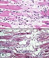
ARVC. (WC)

www
Noncompaction cardiomyopathy
Etiology
- Genetic - LVNC1 gene.[16]
- May be associated with dilation.[16]
- Rare.
- Not clear whether it is a unique entity.[17]
Gross
- Prominent "mesh-like" trabeculae carnae.
- Enlarged intertrabecular recesses.[18]
See also
- Heart.
- Cardiac sarcoidosis.
- Amyloidosis - covers cardiac amyloidosis.
References
- ↑ Cotran, Ramzi S.; Kumar, Vinay; Fausto, Nelson; Nelso Fausto; Robbins, Stanley L.; Abbas, Abul K. (2005). Robbins and Cotran pathologic basis of disease (7th ed.). St. Louis, Mo: Elsevier Saunders. pp. 601. ISBN 0-7216-0187-1.
- ↑ Luk A, Ahn E, Soor GS, Butany J (March 2009). "Dilated cardiomyopathy: a review". J. Clin. Pathol. 62 (3): 219–25. doi:10.1136/jcp.2008.060731. PMID 19017683.
- ↑ DiMaio, Vincent J.M.; Dana, Suzanna E. (2006). Handbook of Forensic Pathology (2nd ed.). CRC Press. pp. 43. ISBN 978-0849392870.
- ↑ Gojanovic B, Feihl F, Gremion G, Waeber B (February 2007). "[Sudden death in young athletes]" (in German). Praxis (Bern 1994) 96 (6): 189-98. PMID 17330410.
- ↑ JB. 9 June 2011.
- ↑ Cotran, Ramzi S.; Kumar, Vinay; Fausto, Nelson; Nelso Fausto; Robbins, Stanley L.; Abbas, Abul K. (2005). Robbins and Cotran pathologic basis of disease (7th ed.). St. Louis, Mo: Elsevier Saunders. pp. 601-3. ISBN 0-7216-0187-1.
- ↑ DiMaio, Vincent J.M.; Dana, Suzanna E. (2006). Handbook of Forensic Pathology (2nd ed.). CRC Press. pp. 44. ISBN 978-0849392870.
- ↑ CK. 14 October 2010.
- ↑ Reddy, M.; Thatai, D.; Bernal, J.; Pradhan, J.; Afonso, L. (Jul 2008). "Apical hypertrophic cardiomyopathy: potential utility of Strain imaging.". Eur J Echocardiogr 9 (4): 560-2. doi:10.1016/j.euje.2007.02.004. PMID 17392031.
- ↑ DiMaio, Vincent J.M.; Dana, Suzanna E. (2006). Handbook of Forensic Pathology (2nd ed.). CRC Press. pp. 44-5. ISBN 978-0849392870.
- ↑ Cutler, DJ.; Isner, JM.; Bracey, AW.; Hufnagel, CA.; Conrad, PW.; Roberts, WC.; Kerwin, DM.; Weintraub, AM. (Dec 1980). "Hemochromatosis heart disease: an unemphasized cause of potentially reversible restrictive cardiomyopathy.". Am J Med 69 (6): 923-8. PMID 7446557.
- ↑ Sudden cardiac death due to hypertrophic cardiomyopathy can be reduced by pre-participation cardiovascular screening in young athletes. URL: http://eurheartj.oxfordjournals.org/cgi/content/full/27/18/2152. Accessed on: 16 December 2009.
- ↑ http://www.ncbi.nlm.nih.gov/entrez/dispomim.cgi?id=601214
- ↑ 14.0 14.1 URL: http://emedicine.medscape.com/article/1612324-overview.
- ↑ Romero J, Mejia-Lopez E, Manrique C, Lucariello R (2013). "Arrhythmogenic Right Ventricular Cardiomyopathy (ARVC/D): A Systematic Literature Review". Clin Med Insights Cardiol 7: 97–114. doi:10.4137/CMC.S10940. PMC 3667685. PMID 23761986. https://www.ncbi.nlm.nih.gov/pmc/articles/PMC3667685/.
- ↑ 16.0 16.1 Online 'Mendelian Inheritance in Man' (OMIM) 604169
- ↑ Paterick TE, Gerber TC, Pradhan SR, Lindor NM, Tajik AJ (2010). "Left ventricular noncompaction cardiomyopathy: what do we know?". Rev Cardiovasc Med 11 (2): 92–9. PMID 20700091.
- ↑ Chin TK, Perloff JK, Williams RG, Jue K, Mohrmann R (August 1990). "Isolated noncompaction of left ventricular myocardium. A study of eight cases". Circulation 82 (2): 507–13. PMID 2372897.