Difference between revisions of "Thecoma"
Jump to navigation
Jump to search
(redirect) |
|||
| (6 intermediate revisions by the same user not shown) | |||
| Line 1: | Line 1: | ||
{{ Infobox diagnosis | |||
| Name = {{PAGENAME}} | |||
| Image = Thecoma_high_mag.jpg | |||
| Width = | |||
| Caption = Thecoma. [[H&E stain]]. | |||
| Synonyms = | |||
| Micro = bland oval or spindled nuclei, abundant cytoplasm that is pale and vaculolated | |||
| Subtypes = | |||
| LMDDx = [[ovarian fibroma]], fibroma-thecoma | |||
| Stains = | |||
| IHC = alpha-inhibin +ve | |||
| EM = | |||
| Molecular = | |||
| IF = | |||
| Gross = solid yellow mass, usually well-circumscribed | |||
| Grossing = | |||
| Site = [[ovary]] - see ''[[ovarian tumours]]'' | |||
| Assdx = | |||
| Syndromes = | |||
| Clinicalhx = | |||
| Signs = | |||
| Symptoms = | |||
| Prevalence = uncommon | |||
| Bloodwork = | |||
| Rads = | |||
| Endoscopy = | |||
| Prognosis = benign | |||
| Other = | |||
| ClinDDx = other [[ovarian tumours]] | |||
| Tx = | |||
}} | |||
'''Thecoma''' is an [[ovarian tumour|ovarian]] [[Sex cord-stromal tumours|sex-cord stromal tumour]]. | |||
==General== | |||
*Associated with compression & atrophy of ovarian cortex, thought to arise from medulla.<ref name=pmid18164409>{{Cite journal | last1 = Nocito | first1 = AL. | last2 = Sarancone | first2 = S. | last3 = Bacchi | first3 = C. | last4 = Tellez | first4 = T. | title = Ovarian thecoma: clinicopathological analysis of 50 cases. | journal = Ann Diagn Pathol | volume = 12 | issue = 1 | pages = 12-6 | month = Feb | year = 2008 | doi = 10.1016/j.anndiagpath.2007.01.011 | PMID = 18164409 }}</ref> | |||
*Approximately 50% have symptoms related to estrogen secretion.<ref name=pmid16810055/> | |||
**May also be viralizing. | |||
==Gross== | |||
Features: | |||
*Solid yellow mass, usually well-circumscribed.<ref name=Ref_AoGP398>{{Ref AoGP|398}}</ref> | |||
DDx: | |||
*[[Ovarian fibroma]] - white solid mass.<ref name=Ref_AoGP398>{{Ref AoGP|398}}</ref> | |||
*Fibroma-thecoma (fibrothecoma). | |||
==Microscopic== | |||
Features:<ref name=pmid16810055>{{Cite journal | last1 = Roth | first1 = LM. | title = Recent advances in the pathology and classification of ovarian sex cord-stromal tumors. | journal = Int J Gynecol Pathol | volume = 25 | issue = 3 | pages = 199-215 | month = Jul | year = 2006 | doi = 10.1097/01.pgp.0000192271.22289.e6 | PMID = 16810055 }}</ref> | |||
*Nuclei with oval to spindle morphology. | |||
*Abundant cytoplasm that is pale, vaculolated -- '''key feature'''. | |||
DDx: | |||
*[[Ovarian fibroma]]. | |||
*[[Leiomyoma]] - rare. | |||
*Other [[sex cord-stromal tumour]]s. | |||
===Images=== | |||
<gallery> | |||
Image:Thecoma_low_mag.jpg | Thecoma - low mag. (WC) | |||
Image:Thecoma_high_mag.jpg | Thecoma - high mag. (WC) | |||
</gallery> | |||
==IHC== | |||
*Alpha-inhibin +ve (90%+).<ref name=pmid16810055/> | |||
==See also== | |||
*[[Ovarian tumours]]. | |||
*[[Ovarian fibroma]]. | |||
==References== | |||
{{Reflist|2}} | |||
[[Category:Diagnosis]] | [[Category:Diagnosis]] | ||
[[Category:Ovarian tumours]] | |||
Latest revision as of 16:10, 29 November 2015
| Thecoma | |
|---|---|
| Diagnosis in short | |
 Thecoma. H&E stain. | |
|
| |
| LM | bland oval or spindled nuclei, abundant cytoplasm that is pale and vaculolated |
| LM DDx | ovarian fibroma, fibroma-thecoma |
| IHC | alpha-inhibin +ve |
| Gross | solid yellow mass, usually well-circumscribed |
| Site | ovary - see ovarian tumours |
|
| |
| Prevalence | uncommon |
| Prognosis | benign |
| Clin. DDx | other ovarian tumours |
Thecoma is an ovarian sex-cord stromal tumour.
General
- Associated with compression & atrophy of ovarian cortex, thought to arise from medulla.[1]
- Approximately 50% have symptoms related to estrogen secretion.[2]
- May also be viralizing.
Gross
Features:
- Solid yellow mass, usually well-circumscribed.[3]
DDx:
- Ovarian fibroma - white solid mass.[3]
- Fibroma-thecoma (fibrothecoma).
Microscopic
Features:[2]
- Nuclei with oval to spindle morphology.
- Abundant cytoplasm that is pale, vaculolated -- key feature.
DDx:
- Ovarian fibroma.
- Leiomyoma - rare.
- Other sex cord-stromal tumours.
Images
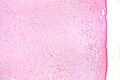
Thecoma - low mag. (WC)
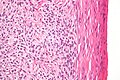
Thecoma - high mag. (WC)

IHC
- Alpha-inhibin +ve (90%+).[2]
See also
References
- ↑ Nocito, AL.; Sarancone, S.; Bacchi, C.; Tellez, T. (Feb 2008). "Ovarian thecoma: clinicopathological analysis of 50 cases.". Ann Diagn Pathol 12 (1): 12-6. doi:10.1016/j.anndiagpath.2007.01.011. PMID 18164409.
- ↑ 2.0 2.1 2.2 Roth, LM. (Jul 2006). "Recent advances in the pathology and classification of ovarian sex cord-stromal tumors.". Int J Gynecol Pathol 25 (3): 199-215. doi:10.1097/01.pgp.0000192271.22289.e6. PMID 16810055.
- ↑ 3.0 3.1 Rose, Alan G. (2008). Atlas of Gross Pathology with Histologic Correlation (1st ed.). Cambridge University Press. pp. 398. ISBN 978-0521868792.