Difference between revisions of "Ovarian fibroma"
Jump to navigation
Jump to search
(split out) |
(fix ref) |
||
| Line 21: | Line 21: | ||
==Microscopic== | ==Microscopic== | ||
Features:<ref>URL: [http://www.pathologyoutlines.com/ovarytumor.html#fibroma http://www.pathologyoutlines.com/ovarytumor.html#fibroma]. Accessed on: 7 May 2012.</ref><ref name=pmid16810055/> | Features:<ref>URL: [http://www.pathologyoutlines.com/ovarytumor.html#fibroma http://www.pathologyoutlines.com/ovarytumor.html#fibroma]. Accessed on: 7 May 2012.</ref><ref name=pmid16810055>{{Cite journal | last1 = Roth | first1 = LM. | title = Recent advances in the pathology and classification of ovarian sex cord-stromal tumors. | journal = Int J Gynecol Pathol | volume = 25 | issue = 3 | pages = 199-215 | month = Jul | year = 2006 | doi = 10.1097/01.pgp.0000192271.22289.e6 | PMID = 16810055 }}</ref> | ||
*[[Spindle cell]]s with central nucleus and no nuclear atypia. | *[[Spindle cell]]s with central nucleus and no nuclear atypia. | ||
*Patternless pattern ([[AKA]] storiform pattern) - not fascicular, not herring bone. | *Patternless pattern ([[AKA]] storiform pattern) - not fascicular, not herring bone. | ||
Revision as of 06:35, 22 October 2014
Ovarian fibroma is a benign ovarian tumour.
General
- May be a part of:
- Meigs syndrome (mnemonic FAR: fibroma, ascites, right pleural effusion).
- Nevoid basal cell carcinoma syndrome (NBCCS), AKA Gorlin syndrome.[1]
- In NBCCS classically - calcified and bilateral.[2]
- Very rarely transform to fibrosarcoma <1%.[3]
Gross
Features:
- Solid white mass, usu. well-circumscribed.[4]
Note:
- Thecoma = yellow solid mass.[4]
Images
www:
Microscopic
- Spindle cells with central nucleus and no nuclear atypia.
- Patternless pattern (AKA storiform pattern) - not fascicular, not herring bone.
- Stainable lipid - minimal or none.[6]
Notes:
- May be cellular.
- Mitotic activity minimal.[7]
DDx:
- Thecoma - lipid.
- Leiomyoma - fascicular architecture, rare in the ovary.
- Fibrosarcoma - nuclear atypia, classically herring bone pattern, very rare.
- Metastatic metaplastic carcinoma - nuclear atypia, rare.
- Endometriosis with extensive fibrosis.
Images
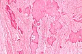
Ovarian fibroma - intermed mag. (WC)
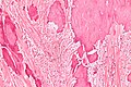
Ovarian fibroma - high mag. (WC)


IHC
- Inhibin -ve (~75%).[6]
Sign out
 OVARIAN MASS ("FIBROMA"), LEFT, EXCISION:
- FIBROMA.
- NEGATIVE FOR MALIGNANCY.
Micro
The sections show spindle cells in a patternless pattern. There is no appreciable nuclear atypia. No mitotic activity is apparent. No necrosis is identified. No calcifications are seen. A small amount of benign ovarian parenchyma is present at the edge of the lesion.
See also
References
- ↑ Cotran, Ramzi S.; Kumar, Vinay; Fausto, Nelson; Nelso Fausto; Robbins, Stanley L.; Abbas, Abul K. (2005). Robbins and Cotran pathologic basis of disease (7th ed.). St. Louis, Mo: Elsevier Saunders. pp. 1103. ISBN 0-7216-0187-1.
- ↑ Tytle, T.; Rosin, D. (Sep 1984). "Bilateral calcified ovarian fibromas.". South Med J 77 (9): 1178-80. PMID 6385289.
- ↑ URL: http://brighamrad.harvard.edu/Cases/bwh/hcache/353/full.html. Accessed on: 4 October 2011.
- ↑ 4.0 4.1 Rose, Alan G. (2008). Atlas of Gross Pathology with Histologic Correlation (1st ed.). Cambridge University Press. pp. 398. ISBN 978-0521868792.
- ↑ URL: http://www.pathologyoutlines.com/ovarytumor.html#fibroma. Accessed on: 7 May 2012.
- ↑ 6.0 6.1 6.2 Roth, LM. (Jul 2006). "Recent advances in the pathology and classification of ovarian sex cord-stromal tumors.". Int J Gynecol Pathol 25 (3): 199-215. doi:10.1097/01.pgp.0000192271.22289.e6. PMID 16810055.
- ↑ Huang, L.; Liao, LM.; Wang, HY.; Zheng, M. (2010). "Clinicopathologic characteristics and prognostic factors of ovarian fibrosarcoma: the results of a multi-center retrospective study.". BMC Cancer 10: 585. doi:10.1186/1471-2407-10-585. PMID 20979607.