Difference between revisions of "Leiomyosarcoma"
Jump to navigation
Jump to search
(+infobox) |
|||
| Line 1: | Line 1: | ||
{{ Infobox diagnosis | |||
| Name = {{PAGENAME}} | |||
| Image = Cutaneous_leiomyosarcoma_-_a_-_intermed_mag.jpg | |||
| Width = | |||
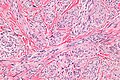
| Caption = Leiomyosarcoma. [[H&E stain]]. | |||
| Micro = | |||
| Subtypes = | |||
| LMDDx = | |||
| Stains = | |||
| IHC = | |||
| EM = | |||
| Molecular = | |||
| IF = | |||
| Gross = | |||
| Grossing = | |||
| Site = [[uterus]], [[skin]] | |||
| Assdx = | |||
| Syndromes = | |||
| Clinicalhx = | |||
| Signs = | |||
| Symptoms = | |||
| Prevalence = uncommon | |||
| Bloodwork = | |||
| Rads = | |||
| Endoscopy = | |||
| Prognosis = poor | |||
| Other = | |||
| ClinDDx = | |||
}} | |||
'''Leiomyosarcoma''' is a malignant tumour of smooth muscle. It is seen in various places including the [[uterus]] and [[skin]]. | '''Leiomyosarcoma''' is a malignant tumour of smooth muscle. It is seen in various places including the [[uterus]] and [[skin]]. | ||
| Line 43: | Line 72: | ||
*[[Endometrial stromal sarcoma]]. | *[[Endometrial stromal sarcoma]]. | ||
Images | ===Images=== | ||
Uterine: | |||
<gallery> | |||
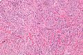
Image:Uterine_leiomyosarcoma_%281%29.jpg | Uterine leiomyosarcoma 1 (WC) | |||
Image:Uterine_leiomyosarcoma_%282%29.jpg | Uterine leiomyosarcoma 2 (WC) | |||
</gallery> | |||
Cutaneous: | |||
<gallery> | |||
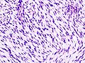
Image:Cutaneous_leiomyosarcoma_-_intermed_mag.jpg | Cutaneous leiomyosarcoma - intermed. mag. - shows fascicular pattern (WC) | |||
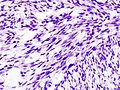
Image:Cutaneous_leiomyosarcoma_-_a_-_intermed_mag.jpg | Cutaneous leiomyosarcoma - intermed. mag. (WC) | |||
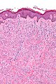
Image:Cutaneous_leiomyosarcoma_-_high_mag.jpg | Cutaneous leiomyosarcoma - high mag. (WC) | |||
</gallery> | |||
www: | |||
*[http://path.upmc.edu/cases/case107/micro.html Cutaneous leiomyosarcoma (upmc.edu)]. | |||
===Subtypes=== | ===Subtypes=== | ||
Major variants:<ref name=Ref_GP284>{{Ref GP|284}}</ref> | Major variants:<ref name=Ref_GP284>{{Ref GP|284}}</ref> | ||
Revision as of 03:57, 29 September 2013
| Leiomyosarcoma | |
|---|---|
| Diagnosis in short | |
 Leiomyosarcoma. H&E stain. | |
| Site | uterus, skin |
|
| |
| Prevalence | uncommon |
| Prognosis | poor |
Leiomyosarcoma is a malignant tumour of smooth muscle. It is seen in various places including the uterus and skin.
General
- Poor prognosis.
- Do not (generally) arise from leiomyomas.
- Often singular, i.e. one tumour; unlike leiomyomas (which are often multiple).
Gross
Features:
- "Fleshy" appearance.
- Necrosis.
- Large size.
- Often singular, i.e. one lesion; leiomyomata are often multiple.
Microscopic
Features:
- Usually a cellular lesion.
- Fasciclar arrangement:
- Whorled look at low power.
- Groups of spindle cells cut peripendicular to their long axis adjacent to groups of spindle cells cut in the plane of their long axis.
- Features of malignancy (usually need 2 of 3): †
- Nuclear atypia.
- Tumour cell necrosis.
- Should be patchy/multifocal.
- Zonal necrosis is suggestive of vascular cause.
- Should be patchy/multifocal.
- Mitoses - key feature - definitions suffer from HPFitis:
- +/-Heterologous elements, e.g. malignant cartilage or bone.[3]
Notes:
- † In deep soft tissue. 1 of 3 criteria is considered enough.[4]
- Leiomyosarcoma de facto trumps other sarcomas.[3]
- Mitotic rate seems to be a relatively weak predictor; modest rate may be malignant and a high rate benign.[5]
DDx:
- Pleomorphic undifferentiated sarcoma.
- EBV-associated smooth muscle tumour - rare, immunoincompetent individuals.
- Carcinosarcoma.
- Smooth muscle tumour of uncertain malignant potential (STUMP).
- Endometrial stromal sarcoma.
Images
Uterine:
Uterine leiomyosarcoma 1 (WC)
Uterine leiomyosarcoma 2 (WC)
.jpg)
.jpg)
Cutaneous:
Cutaneous leiomyosarcoma - intermed. mag. - shows fascicular pattern (WC)
Cutaneous leiomyosarcoma - intermed. mag. (WC)
Cutaneous leiomyosarcoma - high mag. (WC)


www:
Subtypes
Major variants:[6]
- Spindled leiomyosarcoma (leiomyosarcoma NOS) - see above.
- Epithelioid leiomyosarcoma.
- Myxoid leiomyosarcoma.
Minor variants:[6]
- Leiomyosarcoma with prominent intravascular growth.
- Leiomyosarcoma with osteoclast-type cells.
- Leiomyosarcoma with clear cells.
- Leiomyosarcoma with xanthoma-type cells.
Epithelioid leiomyosarcoma
Features:[1]
- >50% epithelial appearance.
- >=5 mitoses/HPF - definition suffers from HPFitis.
Image:
Myxoid leiomyosarcoma
Features:[1]
- >=2 mitoses/HPF - definition suffers from HPFitis.
- May have minimal nuclear atypia.
IHC
Features:
- Positive for SMC markers.
- Desmin - present in all three types of muscle.
- H-caldesmon.
- Smooth muscle myosin.
- CD10 -ve.
- May be +ve.[6]
- Some use in the context of uterine lesions -- CD10 +ve in endometrial stromal sarcoma.
Others:[6]
- ER, PR, AR +ve -- 30-40% of the time.
- CD117 +ve/-ve.
- p53 +ve.
- MIB1 high.
- Kertins usu. -ve -- more often +ve in epithelioid variant.
See also
References
- ↑ 1.0 1.1 1.2 1.3 1.4 Nucci, Marisa R.; Oliva, Esther (2009). Gynecologic Pathology: A Volume in Foundations in Diagnostic Pathology Series (1st ed.). Churchill Livingstone. pp. 281. ISBN 978-0443069208.
- ↑ Kraft S, Fletcher CD (April 2011). "Atypical intradermal smooth muscle neoplasms: clinicopathologic analysis of 84 cases and a reappraisal of cutaneous "leiomyosarcoma"". Am. J. Surg. Pathol. 35 (4): 599–607. doi:10.1097/PAS.0b013e31820e6093. PMID 21358302.
- ↑ 3.0 3.1 Anh Tran, T.; Holloway, RW. (Sep 2012). "Metastatic leiomyosarcoma of the uterus with heterologous differentiation to malignant mesenchymoma.". Int J Gynecol Pathol 31 (5): 453-7. doi:10.1097/PGP.0b013e318246977d. PMID 22833086.
- ↑ URL: http://surgpathcriteria.stanford.edu/softsmoothmuscle/soft_tissue_leiomyosarcoma/differentialdiagnosis.html. Accessed on: 10 May 2013.
- ↑ Guo, L.; Liu, T.; Huang, H. (Oct 1996). "[Reappraisal of the pathological criteria for uterine leiomyosarcoma].". Zhonghua Bing Li Xue Za Zhi 25 (5): 266-9. PMID 9388868.
- ↑ 6.0 6.1 6.2 6.3 Nucci, Marisa R.; Oliva, Esther (2009). Gynecologic Pathology: A Volume in Foundations in Diagnostic Pathology Series (1st ed.). Churchill Livingstone. pp. 284. ISBN 978-0443069208.