Difference between revisions of "Nuclear pleomorphism"
Jump to navigation
Jump to search
(→Images: more) |
|||
| Line 10: | Line 10: | ||
===Images=== | ===Images=== | ||
<gallery> | <gallery> | ||
Image:Glioblastoma_with_extreme_nuclear_enlargement_-_high_mag.jpg | Marked nuclear pleomorphism - in a case of glioblastoma - high mag. (WC) | Image:Glioblastoma_with_extreme_nuclear_enlargement_-_high_mag.jpg | Marked nuclear pleomorphism - in a case of glioblastoma - high mag. (WC/Nephron) | ||
Image:Glioblastoma_with_extreme_nuclear_enlargement_-_very_high_mag.jpg | Marked nuclear pleomorphism - in a case of glioblastoma - very high mag. (WC) | Image:Glioblastoma_with_extreme_nuclear_enlargement_-_very_high_mag.jpg | Marked nuclear pleomorphism - in a case of [[glioblastoma]] - very high mag. (WC/Nephron) | ||
</gallery> | |||
<gallery> | |||
Image:Pleomorphic_undifferentiated_sarcoma_-_intermed_mag.jpg | [[Pleomorphic undifferentiated sarcoma]] - intermed. mag. (WC/Nephron) | |||
Image:Pleomorphic_undifferentiated_sarcoma_-_very_high_mag.jpg | [[Pleomorphic undifferentiated sarcoma]] - high mag. (WC/Nephron) | |||
</gallery> | </gallery> | ||
Revision as of 22:11, 25 September 2013
Nuclear pleomorphism is a common finding in malignant lesions.
It is the marked variation of:[1]
- Nuclear size.
- Nuclear shape.
- Nuclear staining (esp. hyperchromasia).
Memory device 3 S: size, shape, staining.
Images
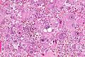
Marked nuclear pleomorphism - in a case of glioblastoma - high mag. (WC/Nephron)
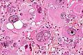
Marked nuclear pleomorphism - in a case of glioblastoma - very high mag. (WC/Nephron)
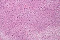
Pleomorphic undifferentiated sarcoma - intermed. mag. (WC/Nephron)
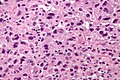
Pleomorphic undifferentiated sarcoma - high mag. (WC/Nephron)


See also
References
- ↑ Rashid, F.; Ul Haque, A. (Dec 2011). "Frequencies of different nuclear morphological features in prostate adenocarcinoma.". Ann Diagn Pathol 15 (6): 414-21. doi:10.1016/j.anndiagpath.2011.06.002. PMID 21849255.